. 2010 Dec 2;116(23):4948-57.
doi: 10.1182/blood-2009-10-247361. Epub 2010 Aug 30.

Transformation by Tribbles homolog 2 (Trib2) requires both the Trib2 kinase domain and COP1 binding

Affiliations

Transformation by Tribbles homolog 2 (Trib2) requires both the Trib2 kinase domain and COP1 binding

Karen Keeshan et al. Blood. .

Abstract

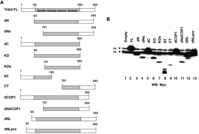
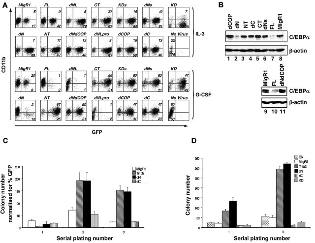
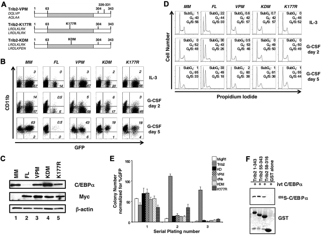
Tribbles homolog 2 (Trib2) is a pseudokinase that induces acute myelogenous leukemia (AML) in mice and is highly expressed in a subset of human AML. Trib2 has 3 distinct regions, a proline-rich N-terminus, a serine/threonine kinase homology domain, and a C-terminal constitutive photomorphogenesis 1 (COP1)-binding domain. We performed a structure-function analysis of Trib2 using in vitro and in vivo assays. The N-terminus was not required for Trib2-induced AML. Deletion or mutation of the COP1-binding site abrogated the ability of Trib2 to degrade CCAAT/enhancer-binding protein-α (C/EBP-α), block granulocytic differentiation, and to induce AML in vivo. Furthermore, COP1 knockdown inhibited the ability of Trib2 to degrade C/EBP-α, showing that it is important for mediating Trib2 activity. We also show that the Trib2 kinase domain is essential for its function. Trib2 contains variant catalytic loop sequences, compared with conventional kinases, that we show are necessary for Trib2 activity. The kinase domain mutants bind, but cannot efficiently degrade, C/EBP-α. Together, our data demonstrate that Trib2 can bind both COP1 and C/EBP-α, leading to degradation of C/EBP-α. Identification of the functional regions of Trib2 that are essential to its oncogenic role provides the basis for developing inhibitors that will block Trib functions in cancer.


Figures

Figure 1
Figure 1
Trib2 mutant design and expression. (A) Schematic of the Trib2 deletion mutants with the amino acid numbers indicated. (B) Expression of Trib2 mutant proteins. 293T cells were transfected with each myc-tagged mutant, and protein lysates were assessed for protein expression by Western blot and probed with the anti-myc antibody 9E10. ns indicates nonspecific binding by the antibody.
Figure 2
Figure 2
The Trib2 N-terminus is dispensable for WT Trib2 activity. (A) Effect of Trib2 mutants on 32D cell differentiation. 32D cells were transduced with the retroviral vector (MigR1), WT Trib2 (FL), or Trib2 mutants and plated in equal numbers (1 × 10⁵; day 0, 48 hours posttransduction) in IL-3 or G-CSF for 5 days. Transduced cells were identified by the expression of the GFP surrogate marker. CD11b expression was assessed after 5 days in culture. Data are representative of 3 independent experiments. (B) Effect of Trib2 mutants on C/EBP-α expression. Sorted GFP+ 32D cells transduced with the indicated retroviral constructs that had been cultured in IL-3 for 2 days were assessed for C/EBP-α protein expression by Western blot. β-actin is the protein loading control. (C) Serial replating ability of Trib2-transduced BM cells. 5-FU–treated BM cells transduced with the retroviral vector (MigR1) or Trib2 constructs were plated in equal number (25 000 unsorted cells in triplicate) in methylcellulose (M3231) containing IL-3, IL-6, SCF, and GM-CSF. The mean numbers of colonies normalized for GFP percentage (as a marker for transduced cells) ± SEM are shown. Data are representative of triplicate cultures from 2 independent experiments. (D) Serial replating ability of Trib2-transduced BM cells from chimeric mice. Then, 25 000 sorted GFP+ BM cells obtained from chimeric mice (MigR1, Trib2, dN, dC, and KD) 6 weeks after BMT and 25 000 control B6 total BM cells were plated in triplicate in methylcellulose (M3434, which contains SCF, IL-3, IL-6, and erythropoietin). The mean numbers of colonies ± SEM are shown. Data are representative of triplicate cultures from 2 independent experiments.
Figure 3
Figure 3
Both the Trib2 KD- and COP1-binding sites are required for Trib2 activity. (A) Schematic of Trib2 point mutants. Left, amino acid sequences show original sequence on top and mutated sequence on bottom. (B) Effect of Trib2 mutants on 32D cell differentiation. 32D cells transduced with the vector control (MigR1-myc; MM), WT Trib2 (FL) or the Trib2 mutants were plated in equal numbers (1 × 10⁵; day 0, 48 hours posttransduction) in IL-3 or G-CSF. Transduced cells were identified by the expression of the GFP surrogate marker. CD11b expression was assessed after 2 and 5 days. Data are representative of 3 independent experiments. (C) Effect of Trib2 mutants on C/EBP-α expression. 32D cells were transduced with the indicated retroviral constructs, sorted for GFP 48 hours after transduction, and assessed for C/EBP-α, myc-tagged FL Trib2, and myc-tagged Trib2 mutant (VPM, KDM, and K177R) protein expression by Western blot, 3 days after sorting. MigR1-Myc is the empty vector (MM). β-actin is the protein loading control. (D) Effect of Trib2 mutants on 32D cell cycle. 32D cells were transduced with the vector control (MigR1-myc; MM), WT Trib2 (FL), or the Trib2 mutants, sorted for GFP 48 hours later, and 1 × 10⁵ GFP+ cells were plated in 5 ng/mL IL-3 or 25 ng/mL G-CSF 3 days after sorting. Cell cycle was assessed by propidium iodide staining on days 2 and 5. (E) Effect of Trib2 mutants on serial replating by transduced BM. 5-FU–treated BM cells transduced with the MigR1 retroviral vector, WT Trib2, or the indicated Trib2 mutants were plated in equal number (25 000 unsorted cells in triplicate) in methylcellulose (M3231), containing IL-3, IL-6, SCF, and GM-CSF. Colonies with > 50 cells were scored and assessed over 3 rounds of serial replating. The mean numbers of colonies normalized for GFP percentage, (as a marker for transduced cells) ± SEM are shown. Data are representative of triplicate cultures from 2 independent experiments. (F) Interaction of Trib2 mutants and C/EBP-α. GST pull-down of Trib2 constructs incubated with IVT 35S C/EBP-α. Top panel: autoradiograph of 35S C/EBP-α. Bottom panel shows Coomassie gel of GST proteins.
Figure 4
Figure 4
The Trib2 COP1-binding site and COP1 are required for C/EBP-α degradation. (A) Interaction of Trib2 and COP1. 293T cells were transfected with COP1-HA (lane 1), myc-tagged Trib2-FL (lane 2), COP1-HA and myc-tagged Trib2-FL (lane 3), myc-tagged dCOP (lane 4), COP1-HA and myc-tagged dCOP (lane 5), myc-tagged VPM (lane 6), COP1-HA and myc-tagged VPM (lane 7), myc-tagged KDM (lane 8), COP1-HA and myc-tagged KDM (lane 9), myc-tagged K177R (lane 10), or COP1-HA and myc-tagged K177R (lane 11). Proteins were immunoprecipitated using the Myc 9E10 antibody, and Western blotting was performed with HA (top panels) and Myc (bottom panels) antibodies on immunoprecipitates (IP), and on whole cell lysates (WCLs). (B) COP1 knockdown decreases COP1 expression. Left panel, 293T cells were transfected with scrambled shRNA, or 1 of 2 different shCOP1 plasmids alone or together with COP1-HA. Western blotting was performed with HA antibody to detect COP1 expression. β-actin served as the loading control. Middle panel, 293T cells were cotransfected with COP1-HA and either the scrambled shRNA or each of the shCOP1 plasmids and analyzed by real-time RT-PCR for COP1 expression. Error bars denote SEM of each sample measured in triplicate. Right panel, sorted GFP+ 32D cells transduced with either shCOP1 or scrambled shRNA-expressing retrovirus were analyzed for endogenous COP1 knockdown by real-time RT-PCR. Error bars denote SEM of each sample measured in triplicate. (C) COP1 knockdown impairs the effects of Trib2 on C/EBP-α expression. Sorted GFP+ tNGFR+ 32D cells transduced with the indicated retroviral constructs were assessed for C/EBP-α protein expression by Western blot. The shRNA constructs were expressed from the low-molecular-mass polypeptide retroviral vector, which expresses GFP as a surrogate marker, whereas Trib2 was expressed from a version of MigR1 that expresses truncated NGFR as a surrogate marker. The cells that were not transduced with FL-Trib2 were transduced with the empty NGFR vector (lanes 2, 4, and 6). β-Actin was the protein loading control.
Figure 5
Figure 5
The COP1-binding site and intact KD are necessary for the leukemogenic activity of Trib2. (A) Kaplan-Meier survival curve of mice receiving Trib2-FL, dN, dCOP, dNs, or MigR1 cotransduced with HoxA9 BM. The median survival of FL and dN mice was 83 and 79 days, respectively. (B) Wright-Giemsa–stained peripheral blood smears from mice receiving full-length Trib2 (FL, top left) and the ΔC (top right), ΔNs (bottom right), and ΔN mice (bottom left). Several leukemic blasts are indicated by the black arrows, and several metamyelocytes are shown by the white arrows. (C) Flow cytometric analysis of BM cells from C57BL/6 mice receiving HoxA9 and/or Trib2 transduced cells. Left panels, analysis of GFP (Trib2 mutants) and HoxA9-NGFR expression (percentages given) in cells obtained from BM of leukemic FL+HoxA9 and dN+HoxA9 mice, compared with C57BL/6 control mice. Flow cytometric analysis of Gr-1 and CD11b expression (middle panels) and CD34 and c-Kit expression (right panels) in the GFP+NGFR+ fractions. Representative plots are shown.

References

    1. Storlazzi CT, Fioretos T, Paulsson K, et al. Identification of a commonly amplified 4.3-Mb region with overexpression of C8FW, but not MYC, in MYC-containing double minutes in myeloid malignancies. Hum Mol Genet. 2004;13(14):1479–1485. - PubMed
    1. Keeshan K, He Y, Wouters BJ, et al. Tribbles homolog 2 inactivates C/EBPalpha and causes acute myelogenous leukemia. Cancer Cell. 2006;10(5):401–411. - PMC - PubMed
    1. Park MH, Cho SA, Yoo KH, et al. Gene expression profile related to prognosis of acute myeloid leukemia. Oncol Rep. 2007;18(6):1395–1402. - PubMed
    1. Jin G, Yamazaki Y, Takuwa M, et al. Trib1 and Evi1 cooperate with Hoxa and Meis1 in myeloid leukemogenesis. Blood. 2007;109(9):3998–4005. - PubMed
    1. Wouters BJ, Jorda MA, Keeshan K, et al. Distinct gene expression profiles of acute myeloid/T-lymphoid leukemia with silenced CEBPA and mutations in NOTCH1. Blood. 2007;110(10):3706–3714. - PMC - PubMed

Publication types

MeSH terms

Substances