

Review
doi: 10.1161/CIRCULATIONAHA.108.847731.
Cell communications in the heart
Affiliations
- PMID: 20805439
- PMCID: PMC2941440
- DOI: 10.1161/CIRCULATIONAHA.108.847731
Item in Clipboard
Review
Cell communications in the heart
Circulation.
.
No abstract available
Conflict of interest statement
Conflict of interest: None
Figures
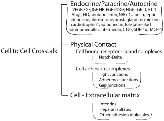
Schematic representation of cell-cell communication modalities in the heart. Types of cell-cell cross-talk and their mediators. Abbreviations are the same as in the text.
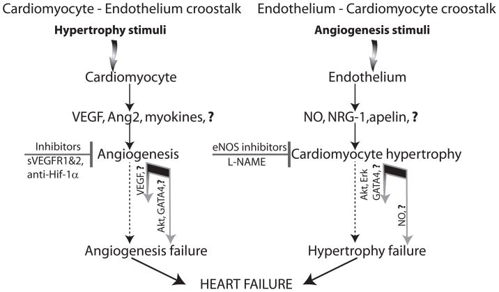
Myocardial hypertrophy as result of cardiomyocyte – endothelium and endothelium – cardiomyocyte crosstalk. Hypertrophic programs can be activated by a direct myocyte stimulation such as pressure or volume overload (left side) or by an expansion of the cardiac vascular mass (right side). Myocyte-driven hypertrophic response includes production of angiogenic growth factors and other myocyte-derived molecules capable of stimulating endothelial expansion (myokines). The angiogenic response is crucial for normal function of the hypertrophic heart and its failure or imbalance in the extent of hypertrophic and angiogenic responses, leads to progressive deterioration of myocardial function and heart failure. On the other hand, increase in the endothelial cell mass in the normal heart can activate myocardial hypertrophy even in the absence of physical stimuli by secretion of nitric oxide, neuregulin (NRG-1) and putative “endokines” (endothelium-derived molecules capable of affecting myocytes). The excess of NO may result in heart failure due to its negative ionotropic properties
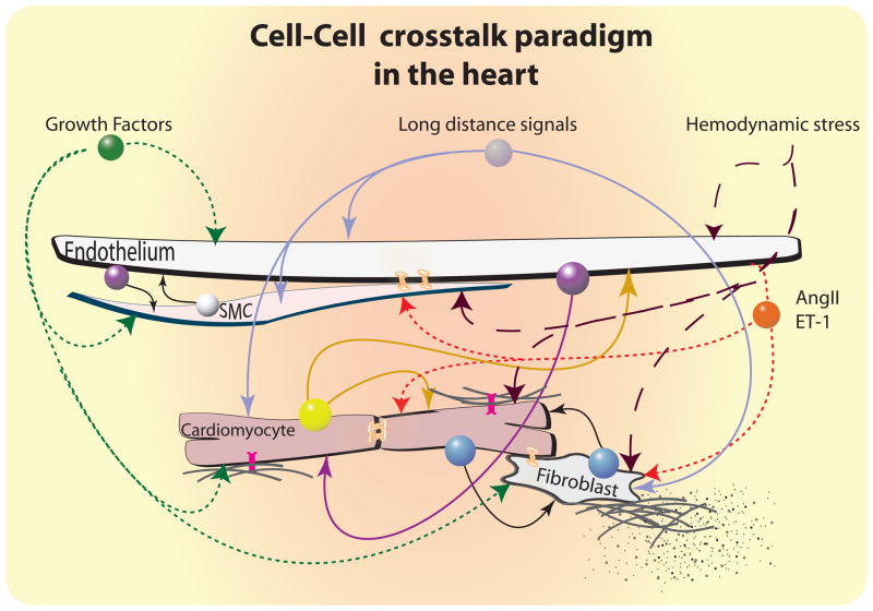
Cellular crosstalk paradigms in the heart. A number of local and long-distance cell-cell communications contribute to maintenance of normal cardiac homeostasis and to responses to hypertrophic stimuli. These include paracrine/autocrine and endocrine signals, direct cell-cell contacts via gap junction, and cell- matrix interactions between cells of the coronary vasculature, cardiomyocytes, fibroblasts and probably other cell types, including tissue-resident stem cells. Perturbations in the balance of this network during sustained hemodynamic stress can lead to a variety of pathological sequelae, including heart failure due to inadequate vascular compensation for myocyte hypertrophy, and the development of cardiac fibrosis as a consequence of fibroblast proliferation and remodeling of the extracellular matrix.  Growth factors (e.g. IGF, FGF, HB-EGF, PDGF, HGF, VEGF, etc.);
  Stress enhancers (e.g. AngII, ET-1, etc.);
  Endothelium signals (e.g. NO, NRG-1, apelin, angiopoientin-1, prostacyclin, etc.);
  SMC (e.g. VEGF, angiopoientin, etc);
  Cardiomyocyte signals (e.g. Fslt-1, VEGF, FGF2, adenosine, etc.);
  Cardiomyocyte-Fibroblast signals (e.g. TGF-β, CTGF, etc.);
  endocrine signals (e.g. adiponectin, adrenomedullin, intermedin, aldosterone etc.);
  integrins;
  connexins.
Growth factors (e.g. IGF, FGF, HB-EGF, PDGF, HGF, VEGF, etc.); Stress enhancers (e.g. AngII, ET-1, etc.); Endothelium signals (e.g. NO, NRG-1, apelin, angiopoientin-1, prostacyclin, etc.); SMC (e.g. VEGF, angiopoientin, etc); Cardiomyocyte signals (e.g. Fslt-1, VEGF, FGF2, adenosine, etc.); Cardiomyocyte-Fibroblast signals (e.g. TGF-β, CTGF, etc.); endocrine signals (e.g. adiponectin, adrenomedullin, intermedin, aldosterone etc.); integrins; connexins.References
-
- Lloyd-Jones D, Adams R, Carnethon M, De Simone G, Ferguson TB, Flegal K, Ford E, Furie K, Go A, Greenlund K, Haase N, Hailpern S, Ho M, Howard V, Kissela B, Kittner S, Lackland D, Lisabeth L, Marelli A, McDermott M, Meigs J, Mozaffarian D, Nichol G, O’Donnell C, Roger V, Rosamond W, Sacco R, Sorlie P, Stafford R, Steinberger J, Thom T, Wasserthiel-Smoller S, Wong N, Wylie-Rosett J, Hong Y. Heart disease and stroke statistics--2009 update: a report from the American Heart Association Statistics Committee and Stroke Statistics Subcommittee. Circulation. 2009;119:480–486. - PubMed
-
- Duncker DJ, Bache RJ. Regulation of coronary blood flow during exercise. Physiol Rev. 2008;88:1009–1086. - PubMed
-
- Hudlicka O, Brown M, Egginton S. Angiogenesis in skeletal and cardiac muscle. Physiol Rev. 1992;72:369–417. - PubMed
-
- Molkentin JD, Dorn GW., 2nd Cytoplasmic signaling pathways that regulate cardiac hypertrophy. Annu Rev Physiol. 2001;63:391–426. - PubMed
Publication types
MeSH terms
Grants and funding
LinkOut - more resources
Full Text Sources
Other Literature Sources
