Refined LexA transactivators and their use in combination with the Drosophila Gal4 system
- PMID: 20805468
- PMCID: PMC2941298
- DOI: 10.1073/pnas.1005957107
Refined LexA transactivators and their use in combination with the Drosophila Gal4 system
Abstract
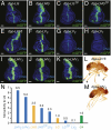
The use of binary transcriptional systems offers many advantages for experimentally manipulating gene activity, as exemplified by the success of the Gal4/UAS system in Drosophila. To expand the number of applications, a second independent transactivator (TA) is desirable. Here, we present the optimization of an additional system based on LexA and show how it can be applied. We developed a series of LexA TAs, selectively suppressible via Gal80, that exhibit high transcriptional activity and low detrimental effects when expressed in vivo. In combination with Gal4, an appropriately selected LexA TA permits to program cells with a distinct balance and independent outputs of the two TAs. We demonstrate how the two systems can be combined for manipulating communicating cell populations, converting transient tissue-specific expression patterns into heritable, constitutive activities, and defining cell territories by intersecting TA expression domains. Finally, we describe a versatile enhancer trap system that allows swapping TA and generating mosaics composed of Gal4 and LexA TA-expressing cells. The optimized LexA system facilitates precise analyses of complex biological phenomena and signaling pathways in Drosophila.
Conflict of interest statement
The authors declare no conflict of interest.
Figures


Similar articles
-
Expanding the Drosophila toolkit for dual control of gene expression.Elife. 2024 Apr 3;12:RP94073. doi: 10.7554/eLife.94073. Elife. 2024. PMID: 38569007 Free PMC article.
-
The GAL4 System: A Versatile System for the Manipulation and Analysis of Gene Expression.Methods Mol Biol. 2016;1478:33-52. doi: 10.1007/978-1-4939-6371-3_2. Methods Mol Biol. 2016. PMID: 27730574 Review.
-
The Q-System: A Versatile Expression System for Drosophila.Methods Mol Biol. 2016;1478:53-78. doi: 10.1007/978-1-4939-6371-3_3. Methods Mol Biol. 2016. PMID: 27730575 Free PMC article. Review.
-
A toolkit for converting Gal4 into LexA and Flippase transgenes in Drosophila.G3 (Bethesda). 2023 Mar 9;13(3):jkad003. doi: 10.1093/g3journal/jkad003. G3 (Bethesda). 2023. PMID: 36617215 Free PMC article.
-
Facilitating Neuron-Specific Genetic Manipulations in Drosophila melanogaster Using a Split GAL4 Repressor.Genetics. 2017 Jun;206(2):775-784. doi: 10.1534/genetics.116.199687. Epub 2017 Mar 31. Genetics. 2017. PMID: 28363977 Free PMC article.
Cited by
-
Imaginal Disc Regeneration: Something Old, Something New.Cold Spring Harb Perspect Biol. 2022 Nov 1;14(11):a040733. doi: 10.1101/cshperspect.a040733. Cold Spring Harb Perspect Biol. 2022. PMID: 34872971 Free PMC article. Review.
-
Mosaic Analysis in Drosophila.Genetics. 2018 Feb;208(2):473-490. doi: 10.1534/genetics.117.300256. Genetics. 2018. PMID: 29378809 Free PMC article. Review.
-
Cytoneme-mediated contact-dependent transport of the Drosophila decapentaplegic signaling protein.Science. 2014 Feb 21;343(6173):1244624. doi: 10.1126/science.1244624. Epub 2014 Jan 2. Science. 2014. PMID: 24385607 Free PMC article.
-
The Little Fly that Could: Wizardry and Artistry of Drosophila Genomics.Genes (Basel). 2014 May 13;5(2):385-414. doi: 10.3390/genes5020385. Genes (Basel). 2014. PMID: 24827974 Free PMC article.
-
Glucocerebrosidase deficiency leads to neuropathology via cellular immune activation.PLoS Genet. 2024 Nov 11;20(11):e1011105. doi: 10.1371/journal.pgen.1011105. eCollection 2024 Nov. PLoS Genet. 2024. PMID: 39527642 Free PMC article.
References
-
- Brand AH, Perrimon N. Targeted gene expression as a means of altering cell fates and generating dominant phenotypes. Development. 1993;118:401–415. - PubMed
-
- Duffy JB. GAL4 system in Drosophila: A fly geneticist's Swiss army knife. Genesis. 2002;34:1–15. - PubMed
-
- Lee T, Luo L. Mosaic analysis with a repressible cell marker for studies of gene function in neuronal morphogenesis. Neuron. 1999;22:451–461. - PubMed
-
- McGuire SE, Le PT, Osborn AJ, Matsumoto K, Davis RL. Spatiotemporal rescue of memory dysfunction in Drosophila. Science. 2003;302:1765–1768. - PubMed
-
- Nellen D, Burke R, Struhl G, Basler K. Direct and long-range action of a DPP morphogen gradient. Cell. 1996;85:357–368. - PubMed
Publication types
MeSH terms
Substances
LinkOut - more resources
Full Text Sources
Other Literature Sources
Molecular Biology Databases

