. 2010 Sep 14;107(37):16291-6.
doi: 10.1073/pnas.1011105107. Epub 2010 Aug 30.

Membrane-shaping host reticulon proteins play crucial roles in viral RNA replication compartment formation and function

Affiliations

Membrane-shaping host reticulon proteins play crucial roles in viral RNA replication compartment formation and function

Arturo Diaz et al. Proc Natl Acad Sci U S A. .

Abstract

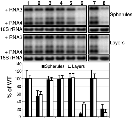
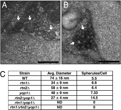
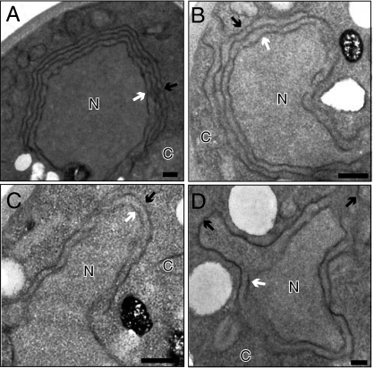
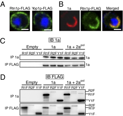
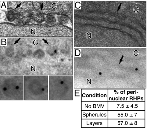
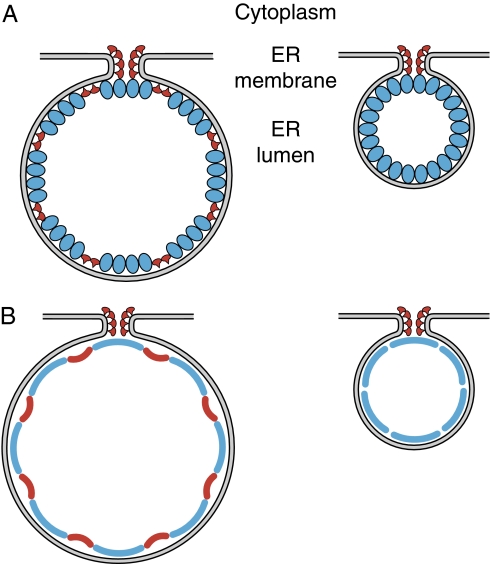
Positive-strand RNA viruses replicate their genomes on membranes with virus-induced rearrangements such as single- or double-membrane vesicles, but the mechanisms of such rearrangements, including the role of host proteins, are poorly understood. Brome mosaic virus (BMV) RNA synthesis occurs in ≈70 nm, negatively curved endoplasmic reticulum (ER) membrane invaginations induced by multifunctional BMV protein 1a. We show that BMV RNA replication is inhibited 80-90% by deleting the reticulon homology proteins (RHPs), a family of membrane-shaping proteins that normally induce and stabilize positively curved peripheral ER membrane tubules. In RHP-depleted cells, 1a localized normally to perinuclear ER membranes and recruited the BMV 2a(pol) polymerase. However, 1a failed to induce ER replication compartments or to recruit viral RNA templates. Partial RHP depletion allowed formation of functional replication vesicles but reduced their diameter by 30-50%. RHPs coimmunoprecipitated with 1a and 1a expression redirected >50% of RHPs from peripheral ER tubules to the interior of BMV-induced RNA replication compartments on perinuclear ER. Moreover, RHP-GFP fusions retained 1a interaction but shifted 1a-induced membrane rearrangements from normal vesicles to double membrane layers, a phenotype also induced by excess 1a-interacting 2a(pol). Thus, RHPs interact with 1a, are incorporated into RNA replication compartments, and are required for multiple 1a functions in replication compartment formation and function. The results suggest possible RHP roles in the bodies and necks of replication vesicles.


Conflict of interest statement

The authors declare no conflict of interest.

Figures

Fig. 1.
Fig. 1.
BMV RNA replication is inhibited in specific reticulon deletion yeast strains. Total RNA extracts were obtained from wt YPH500 (lane 1), rtn1Δ (lane 2), rtn2Δ (lane 3), yop1Δ (lane 4), rtn2Δyop1Δ (lane 5), rtn1Δyop1Δ (lane 6), wt BY4741 (lane 7), or rtn1Δrtn2Δyop1Δ (lane 8) yeast coexpressing 1a, RNA3, and either ADH1 2apol (Spherules) or GAL1 2apol (Layers), and accumulation of RNA3 and subgenomic RNA4 was detected by Northern blotting. Equal loading was verified by probing for 18S ribosomal RNA. Values represent the mean of three independent experiments.
Fig. 2.
Fig. 2.
BMV-induced spherule formation is abolished in the absence of RHPs. Representative EM images of wt (A) and rtn1Δ (B) yeast cells expressing BMV-induced spherules. White arrows point out individual spherular structures. N, nucleus; C, cytoplasm. (Scale bars: 200 nm.) (C) Effect of RHP deletion on spherule diameter and number of spherules per cell.
Fig. 3.
Fig. 3.
Morphology and number of double-membrane layers is altered in triple and one double deletion strain. EM images of yeast cells expressing BMV induced layers in wt (A), rtn1Δ (B), rtn1Δyop1Δ (C), or rtn1Δrtn2Δyop1Δ (D) yeast. Black arrows point out double-membrane layers, whereas white arrows point out the nuclear membrane. N, nucleus; C, cytoplasm. (Scale bars: 200 nm.)
Fig. 4.
Fig. 4.
RHPs interact with BMV 1a protein and relocalize to site of BMV replication. Localization of FLAG-tagged RHPs in yeast transformed with empty plasmids (A) or 1a alone (B). DNA was stained with DAPI (blue). Images were cropped just outside the yeast cell wall to include only one cell. (Scale bars: 2 μm.) (C and D) Cells expressing the FLAG-tagged RHPs and either empty plasmids, 1a alone, or 1a plus GAL1 2apol were lysed, and the cleared lysates were subjected to immunoprecipitation by using anti-1a or anti-FLAG antibodies (IP 1a and IP FLAG, respectively). The resulting immunoprecipitates were analyzed on 4–15% SDS/PAGE and immunoblotted using anti-1a (C) or anti-FLAG (D) antibodies. The positions of 1a, Rtn1p-FLAG (R1F), Rtn2p-FLAG (R2F), and Yop1p-FLAG (Y1F) are shown on the right.
Fig. 5.
Fig. 5.
RHPs localize to interior of spherules and layers. EM images of osmium fixed yeast cells containing spherules (A) or layers (C). Anti-FLAG immunogold EM labeling in yeast cells expressing Rtn1p-FLAG and 1a (B) or 1a plus GAL1 2apol (D). Insets in B show individual spherules. To preserve the antigenicity of the proteins, osmium fixation and staining were reduced, resulting in electron lucent regions where the lipid membranes used to be due to lipid extraction during the dehydration steps. N, nucleus; C, cytoplasm. (E) Percentage of total gold particles that localized to perinuclear ER in yeast expressing no BMV components, spherules, or layers.
Fig. 6.
Fig. 6.
Models for the potential role of RHPs in the formation and/or maintenance of BMV-induced spherules. (A) Schematic of BMV spherules showing how 1a (blue) might induce and stabilize negative membrane curvature in the vesicle body, whereas arced RHP multimers (red) might stabilize the positively curved neck region and also be incorporated in the main vesicle body. (B) Schematics replacing 1a and RHP clusters in the main vesicle body with blue and red arcs, respectively, to show how expansion of the vesicle body in RHP-containing spherule A is primarily due to the action of RHPs in partially neutralizing the intrinsic rate of membrane curvature by 1a alone.

References

    1. Miller S, Krijnse-Locker J. Modification of intracellular membrane structures for virus replication. Nat Rev Microbiol. 2008;6:363–374. - PMC - PubMed
    1. Knoops K, et al. SARS-coronavirus replication is supported by a reticulovesicular network of modified endoplasmic reticulum. PLoS Biol. 2008;6:e226. - PMC - PubMed
    1. Kopek BG, Perkins G, Miller DJ, Ellisman MH, Ahlquist P. Three-dimensional analysis of a viral RNA replication complex reveals a virus-induced mini-organelle. PLoS Biol. 2007;5:e220. - PMC - PubMed
    1. Ahola T, Ahlquist P. Putative RNA capping activities encoded by brome mosaic virus: Methylation and covalent binding of guanylate by replicase protein 1a. J Virol. 1999;73:10061–10069. - PMC - PubMed
    1. Ahola T, den Boon JA, Ahlquist P. Helicase and capping enzyme active site mutations in brome mosaic virus protein 1a cause defects in template recruitment, negative-strand RNA synthesis, and viral RNA capping. J Virol. 2000;74:8803–8811. - PMC - PubMed

Publication types

MeSH terms