

Imaging mass spectrometry of intraspecies metabolic exchange revealed the cannibalistic factors of Bacillus subtilis
- PMID: 20805502
- PMCID: PMC2941286
- DOI: 10.1073/pnas.1008368107
Imaging mass spectrometry of intraspecies metabolic exchange revealed the cannibalistic factors of Bacillus subtilis
Abstract
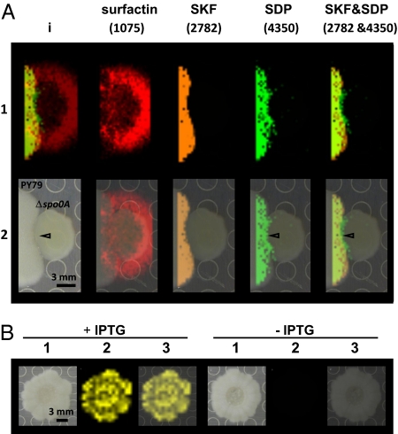
During bacterial cannibalism, a differentiated subpopulation harvests nutrients from their genetically identical siblings to allow continued growth in nutrient-limited conditions. Hypothesis-driven imaging mass spectrometry (IMS) was used to identify metabolites active in a Bacillus subtilis cannibalism system in which sporulating cells lyse nonsporulating siblings. Two candidate molecules with sequences matching the products of skfA and sdpC, genes for the proposed cannibalistic factors sporulation killing factor (SKF) and sporulation delaying protein (SDP), respectively, were identified and the structures of the final products elucidated. SKF is a cyclic 26-amino acid (aa) peptide that is posttranslationally modified with one disulfide and one cysteine thioether bridged to the α-position of a methionine, a posttranslational modification not previously described in biology. SDP is a 42-residue peptide with one disulfide bridge. In spot test assays on solid medium, overproduced SKF and SDP enact a cannibalistic killing effect with SDP having higher potency. However, only purified SDP affected B. subtilis cells in liquid media in fluorescence microscopy and growth assays. Specifically, SDP treatment delayed growth in a concentration-dependent manner, caused increases in cell permeability, and ultimately caused cell lysis accompanied by the production of membrane tubules and spheres. Similarly, SDP but not SKF was able to inhibit the growth of the pathogens Staphylococcus aureus and Staphylococcus epidermidis with comparable IC(50) to vancomycin. This investigation, with the identification of SKF and SDP structures, highlights the strength of IMS in investigations of metabolic exchange of microbial colonies and also demonstrates IMS as a promising approach to discover novel biologically active molecules.
Conflict of interest statement
The authors declare no conflict of interest.
Figures


 Indicates 20 μg/mL;
  . 10 μg/mL;
  , 5 μg/mL;
  , 2 μg/mL;
  , 0.2 μg/mL; and
  , DMSO control. (B) Growth curves of KP648 (Δspo0A) in ISP2 media with 20 μg/mL SDP. SDP was added at 3 h (
 ) and 6 h (
 ).
). Indicates DMSO control. (C) Fluorescence micrographs of growing cells of 3610, PY79, ALB1035 (3610, Δspo0A), and KP648 (PY79, Δspo0A) treated with DMSO or 20 μg/mL SDP for the time indicated. Red stain is FM 4–64, a fluorescent membrane stain; blue and green stains are DAPI and Sytox Green, two DNA stains that are membrane impermeable. Sytox Green is the least permeable and provides the greatest increase in fluorescence in permeabilized cells. White arrows point to dividing cells. In the DMSO control, the arrow points to a normal division, whereas in the other images the septa are asymmetric. Double arrowheads point to large gaps in membrane staining. Light blue triangles point to membrane spheres, whereas white triangles point to tubular membranes. After 300 min of treatment of PY79 with SDP, surviving cells are smaller, dividing, and impermeable to Sytox. (D) Time-lapse microscopy images collected at 110 min after SDP treatment. Still images from 0 to ∼21 s are shown.

References
-
- Kunst F, et al. The complete genome sequence of the gram-positive bacterium Bacillus subtilis. Nature. 1997;390:249–256. - PubMed
Publication types
MeSH terms
Substances
Grants and funding
LinkOut - more resources
Full Text Sources
Other Literature Sources
Molecular Biology Databases
