

doi: 10.1084/jem.20101157.
Find-me and eat-me signals in apoptotic cell clearance: progress and conundrums
Affiliations
- PMID: 20805564
- PMCID: PMC2931173
- DOI: 10.1084/jem.20101157
Item in Clipboard
Find-me and eat-me signals in apoptotic cell clearance: progress and conundrums
J Exp Med.
.
Abstract
Everyday we turnover billions of cells. The quick, efficient, and immunologically silent disposal of the dying cells requires a coordinated orchestration of multiple steps, through which phagocytes selectively recognize and engulf apoptotic cells. Recent studies have suggested an important role for soluble mediators released by apoptotic cells that attract phagocytes ("find-me" signals). New information has also emerged on multiple receptors that can recognize phosphatidylserine, the key "eat-me" signal exposed on the surface of apoptotic cells. This perspective discusses recent exciting progress, gaps in our understanding, and the conflicting issues that arise from the newly acquired knowledge.
Figures
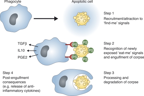
Different steps involved in efficient apoptotic cell clearance. The find-me signals (such as low levels of nucleotides ATP and UTP, fractalkine, lysophosphatidylcholine, or sphingosine 1-phosphate) released by apoptotic cells help attract motile phagocytes to the proximity of the cell undergoing apoptosis. The phagocytes then use engulfment receptors on their surface to engage eat-me signals on apoptotic cells. For clarity, only the PtdSer on the apoptotic cells engaged by cognate receptors is depicted. Engagement of the engulfment receptors (linked to PtdSer recognition) has been shown to stimulate release of antiinflammatory cytokines such as TGF-β, IL-10, and prostaglandin E2 (PGE2). The intracellular signaling induced within the phagocyte by the ligand–receptor interactions leads to cytoskeletal rearrangements and internalization of the dying cell. The phagocyte processes the engulfed corpse through a series of steps, and proper digestion seems to be important for continued uptake of other dying cells by phagocytes.
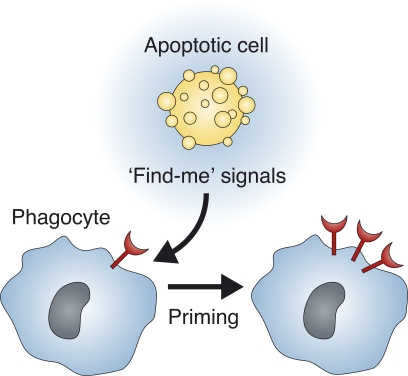
Potential effect of find-me signals on phagocytes. Find-me signals may prime phagocytes to up-regulate the engulfment machinery or cause an increase in expression of engulfment receptors (red) or other components that contribute to engulfment (such as cytoplasmic signaling proteins or soluble bridging molecules; not depicted).
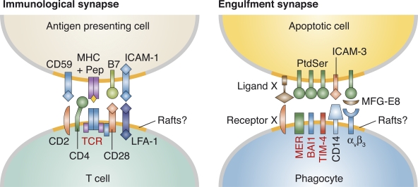
A possible engulfment synapse during apoptotic cell clearance. In a speculative model, an engulfment synapse may help phagocyte binding to and subsequent internalization of apoptotic target cells. There appears to be some clear similarities between T cell–APC recognition and phagocyte–apoptotic cell interactions (see text for more details). However, whereas the T cell–APC interaction is anchored by MHC+peptide interaction with the TCR, the PtdSer exposed on apoptotic cells perhaps may be more homogeneous (except for modifications such as oxidation, etc.), and is likely recognized by multiple PtdSer recognition receptors on the phagocyte. How the PtdSer is displayed on the apoptotic cells and how the different engulfment receptors are arranged on the phagocyte are not yet defined. One could speculate that a second signal analogous to B7 (ligand X) on the apoptotic cell may engage a co-stimulatory receptor analogous to CD28 (receptor X) on the phagocyte, thereby providing specificity, overcoming don’t-eat-me signals, lowering the threshold for phagocyte priming, and/or altering membrane curvature.
References
Publication types
MeSH terms
Substances
Grants and funding
LinkOut - more resources
Full Text Sources
Other Literature Sources
