

The role of crumbs genes in the vertebrate cornea
- PMID: 20805571
- PMCID: PMC2941173
- DOI: 10.1167/iovs.09-4549
The role of crumbs genes in the vertebrate cornea
Abstract
Purpose: To evaluate the role of crumbs genes and related epithelial polarity loci in the vertebrate cornea.
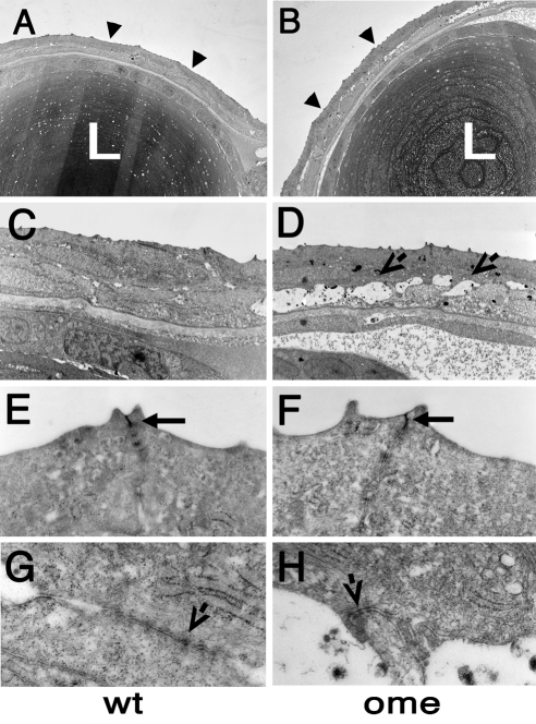
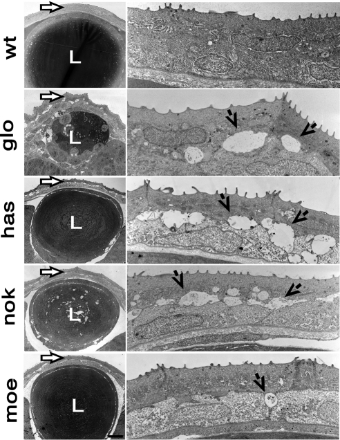
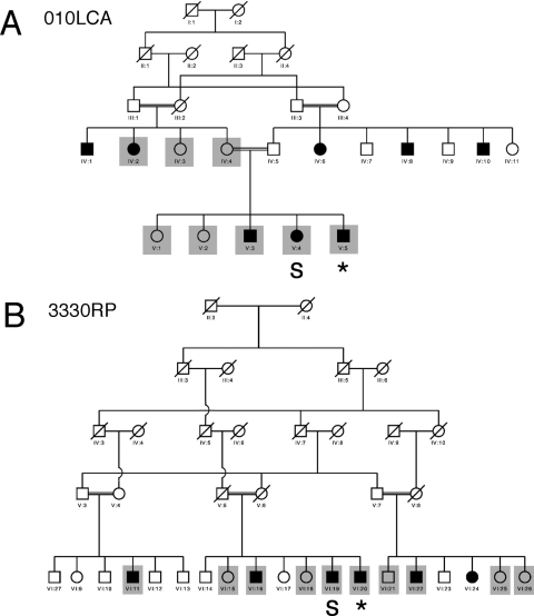
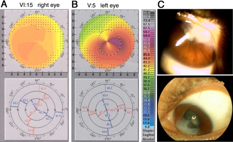
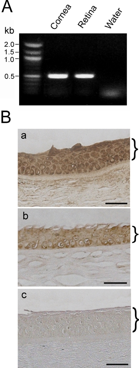
Methods: The authors used histologic analysis and electron microscopy to evaluate the corneas of zebrafish mutant for a crumbs locus oko meduzy (ome) and in mutants of four other loci, nagie oko (nok), heart and soul (has), mosaic eyes (moe), and ncad (formerly glass onion), that function in the same or related genetic pathways. In parallel, they performed an evaluation of corneas in human carriers of a crumbs gene, CRB1, and mutations using topography and biomicroscopy. The expression of the CRB1 gene in the normal human cornea was examined by polymerase chain reaction (PCR) and immunohistochemical staining.
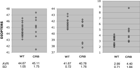
Results: The corneas of zebrafish mutants display severe abnormalities of the epithelial and stromal layers. The epithelial cells do not properly adhere to each other, and fluid-filled spaces form between them. In addition, the layering of the corneal stroma is poorly formed or absent. The corneas of human carriers of CRB1 mutations display shape deviations compared with what has been observed in normal individuals. A PCR product of the correct size was obtained from normal human corneal samples. Sequence analyses confirmed its identity to be the human CRB1 gene. Immunohistochemical staining using anti-CRB1 yielded positive brown deposits in the human cornea.
Conclusions: crumbs genes play a role in the differentiation of the vertebrate cornea. Corneal defects associated with crumbs gene mutations are very severe in the zebrafish model and, in comparison, appear clinically less pronounced in the human eye.
Figures
References
-
- McMahon TT, Szczotka-Flynn L, Barr JT, et al. A new method for grading the severity of keratoconus: the Keratoconus Severity Score (KSS). Cornea. 2006;25:794–800 - PubMed
-
- Poulaki V, Colby K. Genetics of anterior and stromal corneal dystrophies. Semin Ophthalmol. 2008;23:9–17 - PubMed
-
- Javadi MA, Rafee'i AB, Kamalian N, Karimian F, Ja'farinasab MR, Yazdani S. Concomitant keratoconus and macular corneal dystrophy. Cornea. 2004;23:508–512 - PubMed
-
- Kennedy RH, Bourne WM, Dyer JA. A 48-year clinical and epidemiologic study of keratoconus. Am J Ophthalmol. 1986;101:267–273 - PubMed
-
- Hofstetter HW. A keratoscopic survey of 13,395 eyes. Am J Optom Arch Am Acad Optom. 1959;36:3–11 - PubMed
Publication types
MeSH terms
Substances
Grants and funding
- EY01792/EY/NEI NIH HHS/United States
- EY018728/EY/NEI NIH HHS/United States
- EY03890/EY/NEI NIH HHS/United States
- R21 EY018728/EY/NEI NIH HHS/United States
- EY016859/EY/NEI NIH HHS/United States
- R01 EY016859/EY/NEI NIH HHS/United States
- R01 EY003890/EY/NEI NIH HHS/United States
- 063406/Z/2000/Z/WT_/Wellcome Trust/United Kingdom
- R03 EY017571/EY/NEI NIH HHS/United States
- P30 EY001792/EY/NEI NIH HHS/United States
- EY017571/EY/NEI NIH HHS/United States
- EY10608/EY/NEI NIH HHS/United States
- R01 EY011882/EY/NEI NIH HHS/United States
- P30 EY010608/EY/NEI NIH HHS/United States
- WT_/Wellcome Trust/United Kingdom
LinkOut - more resources
Full Text Sources
Molecular Biology Databases
Research Materials
