. 2010 Sep;136(3):293-309.
doi: 10.1085/jgp.201010480.

Dual roles of the sixth transmembrane segment of the CFTR chloride channel in gating and permeation

Affiliations

Dual roles of the sixth transmembrane segment of the CFTR chloride channel in gating and permeation

Yonghong Bai et al. J Gen Physiol. 2010 Sep.

Abstract

Cystic fibrosis transmembrane conductance regulator (CFTR) is the only member of the adenosine triphosphate-binding cassette (ABC) transporter superfamily that functions as a chloride channel. Previous work has suggested that the external side of the sixth transmembrane segment (TM6) plays an important role in governing chloride permeation, but the function of the internal side remains relatively obscure. Here, on a cysless background, we performed cysteine-scanning mutagenesis and modification to screen the entire TM6 with intracellularly applied thiol-specific methanethiosulfonate reagents. Single-channel amplitude was reduced in seven cysteine-substituted mutants, suggesting a role of these residues in maintaining the pore structure for normal ion permeation. The reactivity pattern of differently charged reagents suggests that the cytoplasmic part of TM6 assumes a secondary structure of an alpha helix, and that reactive sites (341, 344, 345, 348, 352, and 353) reside in two neighboring faces of the helix. Although, as expected, modification by negatively charged reagents inhibits anion permeation, interestingly, modification by positively charged reagents of cysteine thiolates on one face (344, 348, and 352) of the helix affects gating. For I344C and M348C, the open time was prolonged and the closed time was shortened after modification, suggesting that depositions of positive charges at these positions stabilize the open state but destabilize the closed state. For R352C, which exhibited reduced single-channel amplitude, modifications by two positively charged reagents with different chemical properties completely restored the single-channel amplitude but had distinct effects on both the open time and the closed time. These results corroborate the idea that a helix rotation of TM6, which has been proposed to be part of the molecular motions during transport cycles in other ABC transporters, is associated with gating of the CFTR pore.


Figures
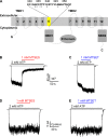
Figure 1.
Figure 1.
Cysless CFTR is an appropriate background for cysteine scanning. (A) Topological diagram of CFTR showing its domain architecture: two TMDs, two NBDs, and one R domain. Each TMD contains six TMs (1–6 and 7–12) connected by intracellular and extracellular loops. Cysteines were introduced into TM6 (yellow). The sequence of TM6 is shown, and residues where cysteine-substitution affects single-channel amplitude are indicated by stars. (B and C) Modification of phosphorylated WT CFTR channels by MTSET and MTSES. After the ATP-induced current reached a plateau, the solution was exchanged from ATP to ATP plus MTSES or MTSET using the fast solution exchange system (see Materials and methods). The solution was switched back to ATP after the MTS reagents took full effect. The dotted line represents the baseline in all figures. (D and E) Similar protocols as in B and C showing that cysless CFTR channels do not respond to the treatment of MTS.
Figure 2.
Figure 2.
Effects of cysteine substitutions on single-channel current amplitude. (A) Single-channel traces for cysless/WT and two cysteine-substituted mutants. Single-channel amplitude: cysless/WT, 0.46 ± 0.005 pA (n = 5); cysless/K355C, 0.28 ± 0.011 pA (n = 4); cysless/F337C, 0.19 ± 0.008 pA (n = 3). (B) Summary of the single-channel amplitude for cysless/WT and all mutant channels. The data are shown as mean and SEM of three to seven experiments. Stars indicate a significant difference in the single-channel amplitude from the cysless/WT, as determined by Student’s t test. Daggers indicate channels whose single-channel amplitude is too small and/or variable to be measured accurately.
Figure 3.
Figure 3.
Summary of the change in macroscopic mean currents after modification. The percentage change in the mean current is calculated as (I/I0−1) × 100%, where I0 and I are the mean current before and after modification, respectively. The left column is the effect of MTSES (red), and the right is the effect of MTSET (blue). n = 3–8 for each construct. Stars indicate mutant channels exhibiting a reduced single-channel amplitude.
Figure 4.
Figure 4.
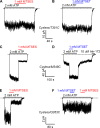
Three different patterns of functional consequences after MTS modification. (A and B) Neither MTSES nor MTSET altered the current of cysless/T351C channels. (C) Modification of 348C by MTSES decreased the current. (D) MTSET increased ATP-dependent current in cysless/M348C channels. After the removal of ATP, there remained a large amount of ATP-independent current, which disappeared upon the application of 10 µM CFTRinh-172. (E) MTSES reduced the current of cysless/Q353C channels. (F) For cysless/Q353C, the treatment of MTSET had little influence on channel function, but prevented the current decrease in response to MTSES, as in E.
Figure 5.
Figure 5.
Effects of MTSET on a single cysless/M348C channel. (A) Single-channel traces in the presence of 2 mM ATP before (left top trace) and after MTSET modification (left middle trace, blue), and after DTT treatment (left bottom trace) in the same patch. Corresponding all-point histograms (gray bars) and their Gaussian fits (black and blue lines) were plot on the right. (B) Gating parameters of the cysless/M348C channel before (black) and after (blue) modification, as extracted from the traces in A. Those of the cysless/WT (gray) in the presence of 2 mM ATP are also included for comparison (traces not depicted). n = 5 for cysless/M348C. n = 6 for cysless/WT. (C) Single-channel amplitude of the cysless/M348C channel before and after modification in the same patch. Star indicates significant difference as determined by Student’s paired t test; n = 5.
Figure 6.
Figure 6.
A single-channel recording of cysless/344C showing the effect of MTSET. (A) Gating behavior in the presence of 2 mM ATP before modification (left top trace), after modification (left middle trace, blue), and after the reversal of the modification by DTT (left bottom trace) in the same patch. Corresponding all-point histograms (gray bars) and their Gaussian fits (black and blue lines) were plot on the right. (B) The Po, obtained from the traces in A, of the cysless/I344C channel before (black) and after (blue) modification. Those of the cysless/WT (gray) in the presence of 2 mM ATP are included for comparison (traces not depicted). n = 7 for cysless/I344C. n = 6 for cysless/WT. (C) Single-channel amplitude of the cysless/I344C channel before and after modification. Star indicates significant difference as determined by Student’s paired t test; n = 7.
Figure 7.
Figure 7.
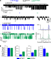
A single-channel recording showing that modification of 352C by MTSET and MTSEA affects both Po and single-channel amplitude. (A; top trace) A continuous recording showing the effects of MTSET or MTSEA on a single cysless/R352C channel. (Bottom traces) Expanded recordings for segments I–III marked in the top trace. A short part in segment I was further zoomed out in a. Corresponding all-points amplitude histograms of II and III were depicted. Blue and green lines are the results of Gaussian fitting. (B) Single-channel amplitude, Po, open time and closed time of MTSET- (blue) and MTSEA-modified (green) cysless/R352C channel, as determined by Gaussian fitting and kinetics analysis; n = 6.
Figure 8.
Figure 8.
Blocking of cysless/R352C channels by glibenclamide before and after MTS modification. (A and B) The blocking effect of 50 µM glibenclamide was enhanced after the channels were modified by MTSET and MTSEA. (C) Percentage change in mean current induced by MTSET (blue) and MTSEA (green). (D) The fraction of block, calculated as (1−Ig/I0) × 100% (Ig and I0 are the mean current in the presence of ATP and ATP plus glibenclamide, respectively), for cysless/WT channels (gray), cysless/R352C channels before modification (black) and after modification with MTSET (blue), and after modification with MTSEA (green). Star indicates a significant difference from cysless/WT channels.
Figure 9.
Figure 9.
Modification of 353C by negatively charged MTSES reduced the single-channel amplitude. This representative recording (among five patches) contains two cysless/Q353C channels. After the application of MTSES, the single-channel amplitude was reduced. Segment a (red) is expanded to show the varying single-channel amplitude after modification.
Figure 10.
Figure 10.
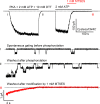
MTSES decreased single-channel amplitude of cysless/I344C channels. (Top trace) A continuous recoding from a patch containing hundreds of channels first phosphorylated with PKA, ATP, and DTT (see Materials and methods) until the current is steady. The current in the presence of ATP was dramatically reduced by the treatment of MTSES. (Bottom traces) Expansions of segments I–III marked in the top trace. Segment I shows spontaneous openings in the absence of ATP. Segments II and III show the current relaxation after the removal of ATP.
Figure 11.
Figure 11.
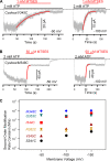
Reaction rates of MTSES modification at various membrane potentials. (A and B) Macroscopic recordings of cysless/V345C and cysless/M348C showing modification by 1 mM MTSES when the membrane potential is held at −50 mV (left) and −100 mV (right). Red lines are the results of exponential fitting. Note that the time scales of the left and the right traces are the same. (C) Summary of the second-order reaction rate constants at three different membrane potentials. n = 4–7 for each data point.
Figure 12.
Figure 12.
(A) Cartoons showing putative helical structures of part of TM6 (341–354). Structure of the cytoplasmic part of TM6 was published by Serohijos et al. (2008). Six reactive sites identified by this study were represented as colored sticks. Position 349, albeit not reactive (indicated by parentheses), was included for clarity. Figures were prepared with PyMOL (V0.99; Schrödinger). (B) A continuous single-channel recording of cysless/I344C showing a dramatic increase of the spontaneous ATP-independent gating after MTSET modification. Before MTSET modification, negligible opening events were observed in the absence of ATP. After MTSET modification, the channel activity without ATP is nearly the same as unmodified channel in the presence of ATP. This phenotype can be reversed by DTT. For spontaneous ATP-independent openings, the mean open time is 0.38 ± 0.02 s (n = 3) before modification (because these openings are rare, we collected events in patches yielding macroscopic currents after ATP was removed as shown in Fig. 10 and performed statistical analysis) and 0.72 ± 0.06 s (n = 5) after modification. Similar results were obtained for cysless/M348C: 0.36 ± 0.03 s (n = 3) before and 0.55 ± 0.03 s (n = 3) after modification. (C) Homology model of CFTR using the structure of Sav1866 as a template (adopted from Serohijos et al., 2008). The coordinate of the homology model was published by Serohijos et al. (2008). Two horizontal lines depict the possible boundaries of the membrane. TMD1 and NBD1 were colored yellow, TMD2 and NBD2 were colored green, and the R domain was colored cyan. Part of TM3 (residues 199–217) was removed to show a clear view of TM6 (residues 330–352, blue). S341 and R352 (red) were represented as sticks.

References

    1. Alexander C., Ivetac A., Liu X., Norimatsu Y., Serrano J.R., Landstrom A., Sansom M., Dawson D.C. 2009. Cystic fibrosis transmembrane conductance regulator: using differential reactivity toward channel-permeant and channel-impermeant thiol-reactive probes to test a molecular model for the pore. Biochemistry. 48:10078–10088 10.1021/bi901314c - DOI - PMC - PubMed
    1. Aubin C.N., Linsdell P. 2006. Positive charges at the intracellular mouth of the pore regulate anion conduction in the CFTR chloride channel. J. Gen. Physiol. 128:535–545 10.1085/jgp.200609516 - DOI - PMC - PubMed
    1. Bear C.E., Li C.H., Kartner N., Bridges R.J., Jensen T.J., Ramjeesingh M., Riordan J.R. 1992. Purification and functional reconstitution of the cystic fibrosis transmembrane conductance regulator (CFTR). Cell. 68:809–818 10.1016/0092-8674(92)90155-6 - DOI - PubMed
    1. Beck E.J., Yang Y., Yaemsiri S., Raghuram V. 2008. Conformational changes in a pore-lining helix coupled to cystic fibrosis transmembrane conductance regulator channel gating. J. Biol. Chem. 283:4957–4966 10.1074/jbc.M702235200 - DOI - PubMed
    1. Bompadre S.G., Ai T., Cho J.H., Wang X., Sohma Y., Li M., Hwang T.C. 2005. CFTR gating I. Characterization of the ATP-dependent gating of a phosphorylation-independent CFTR channel (DeltaR-CFTR). J. Gen. Physiol. 125:361–375 10.1085/jgp.200409227 - DOI - PMC - PubMed

Publication types

MeSH terms

Substances