. 2010 Dec;58(12):1093-106.
doi: 10.1369/jhc.2010.956870. Epub 2010 Aug 30.

Newer insights into premeiotic development of germ cells in adult human testis using Oct-4 as a stem cell marker

Affiliations

Newer insights into premeiotic development of germ cells in adult human testis using Oct-4 as a stem cell marker

Deepa Bhartiya et al. J Histochem Cytochem. 2010 Dec.

Abstract

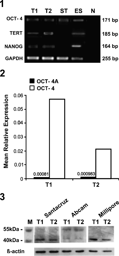
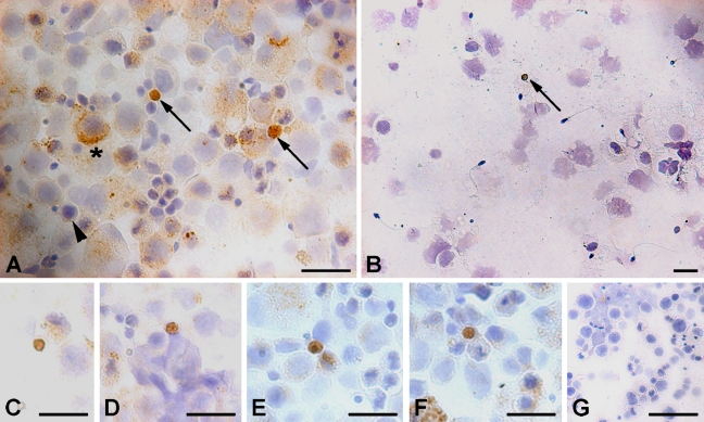
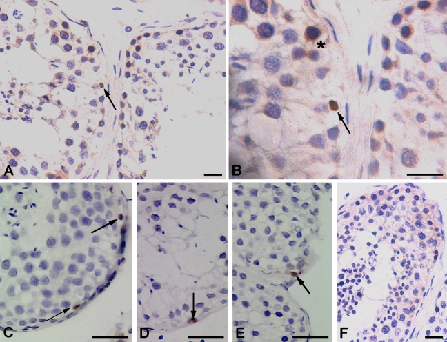
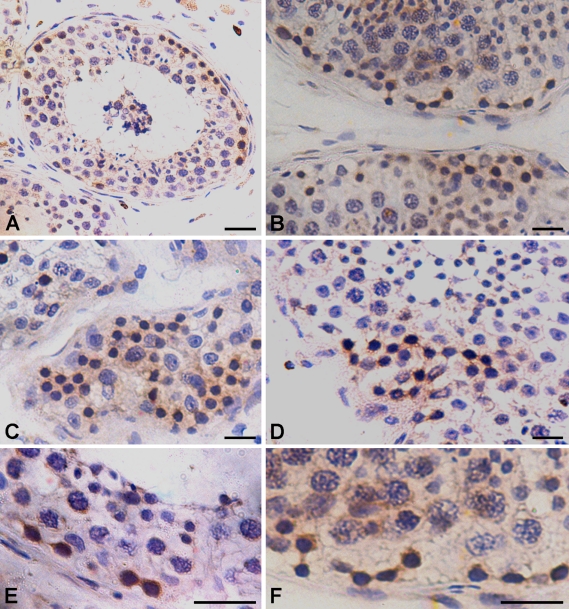
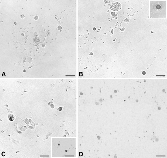
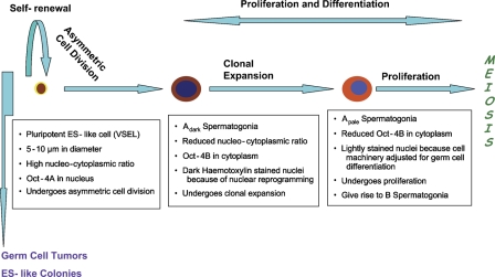
The transcription factor octamer-binding transforming factor 4 (Oct-4) is central to the gene regulatory network responsible for self-renewal, pluripotency, and lineage commitment in embryonic stem (ES) cells and induced pluripotent stem cells (PSCs). This study was undertaken to evaluate differential localization and expression of two major transcripts of Oct-4, viz. Oct-4A and Oct-4B, in adult human testis. A novel population of 5- to 10-μm PSCs with nuclear Oct-4A was identified by ISH and immunolocalization studies. Besides Oct-4, other pluripotent markers like Nanog and TERT were also detected by RT-PCR. A(dark) spermatogonial stem cells (SSCs) were visualized in pairs and chains undergoing clonal expansion and stained positive for cytoplasmic Oct-4B. Quantitative PCR and Western blotting revealed both the transcripts, with higher expression of Oct-4B. It is proposed that PSCs undergo asymmetric cell division and give rise to A(dark) SSCs, which proliferate and initiate lineage-specific differentiation. The darkly stained nuclei in A(dark) SSCs may represent extensive nuclear reprogramming by epigenetic changes when a PSC becomes committed. Oct-4B eventually disappeared in mature germ cells, viz. spermatocytes, spermatids, and sperm. Besides maintaining normal testicular homeostasis, PSCs may also be implicated in germ cell tumors and ES-like colonies that have recently been derived from adult human testicular tissue.


Figures

Figure 1
Figure 1
RT-PCR amplification of pluripotent markers in adult human testicular tissue. Bands of expected sizes (164, 171, 185, and 255 bps) corresponding to NANOG, octamer-binding transforming factor 4 (Oct-4), TERT, and glyceraldehyde-3-phosphate dehydrogenase (GAPDH) were detected in both the testicular samples T1 and T2, in positive control embryonic stem (ES) cells, and also in adult somatic stromal cells (ST) because primers used were common for Oct-4A and Oct-4B. No bands were detected in the negative control (N). However, NANOG and TERT did not amplify in adult somatic stromal cells.
Figure 2
Figure 2
Relative expression of Oct-4 and Oct-4A mRNA by quantitative PCR (Q-PCR). Values are of mean relative expression of two adult human testicular samples T1 and T2 normalized to 18S rRNA by Q-PCR. Expression of Oct-4A isoform transcript was very low compared with that of the total Oct-4 (Oct-4A and Oct-4B) transcript in both the samples analyzed.
Figure 3
Figure 3
Western blot analysis of Oct-4 in adult human testis. An expected band of 43 kDa was detected in both the samples (T1 and T2) using antibodies directed against the N-terminal, specific to Oct-4A isoform (Millipore and Santa Cruz) and antibody directed against the common C-terminus (Abcam). An additional band ∼55 kDa was also observed with the Abcam antibody, as reported earlier in mice (www.abcam.com/ab19857). β-actin was used as the loading control.
Figure 4
Figure 4
Immunocytochemical localization of Oct-4A and Oct-4B in testicular germ cell smears. A distinct cell population stained positive using polyclonal antibody (Abcam) with nuclear Oct-4A and a characteristic unstained rim of cytoplasm (arrow, A). These cells were few in number, thus different fields were captured to demonstrate their presence (C–F). Another population comprised large-sized cells with cytoplasmic Oct-4B (asterisk, A). A distinct gradation of staining intensity for Oct-4B was observed in the cytoplasm of these cells, ranging from dark brown to no stain (arrowhead). Only nuclear Oct-4-positive cells (arrow) were detected using MAb from Santa Cruz (B), and no stain was observed in negative control (G). Bar = 20 μm.
Figure 5
Figure 5
Immunohistochemical localization of Oct-4 pluripotency network in adult human testis. (A,B) Using polyclonal antibody from Abcam, two distinct staining patterns were observed in seminiferous tubules, including cells with nuclear Oct-4A (arrow) and spermatogonial Adark cells with cytoplasmic Oct-4B (asterisk). Three different representative fields of seminiferous tubules show the presence of Oct-4A-positive cells (arrow) using MAb from Santa Cruz (C–E) and negative control showed no staining (F). Bar = 20 μm.
Figure 6
Figure 6
Immunohistochemical localization of cytoplasmic Oct-4B in spermatogonial Adark cells showing extensive proliferation. The cytoplasmic Oct-4B-positive spermatogonial Adark cells are present in patches next to the basement membrane of the seminiferous tubule (A). These cells show proliferation and are easily visualized in pairs (B,E,F) and also in chains (C,D). They undergo clonal expansion and cytoplasmic bridges between dividing cells are easily visualized as they also stain positive for Oct-4B. The cells appear dark and stand out in the section because of brown cytoplasmic Oct-4B and hematoxylin-stained dark nuclei, characteristic of Adark spermatogonial cells. Bar = 20 μm.
Figure 7
Figure 7
Oct-4A mRNA expression on adult human testicular germ cell smears. Different fields show the presence of distinct nuclear staining for Oct-4A mRNA (A–C) in small-sized cells with a distinct unstained cytoplasm. Many different cell types were observed but were distinctly negative for Oct-4A expression. Insets: B, high magnification image of a positively stained cell; C, positively stained cells. The negative control (D) hybridized with sense oligoprobe showed no expression of Oct-4A. Bar = 20 μm.
Figure 8
Figure 8
Revised scheme for premeiotic development of germ cells in adult human testis. It is hypothesized that small and rare ES-like cells detected for the first time in testis undergo asymmetric cell division to give rise to Adark spermatogonial cells with cytoplasmic Oct-4B. These cells undergo extensive nuclear reprogramming by epigenetic changes because pluripotent cell machinery is shut OFF and specific genes necessary for committed cell fate are turned ON. This may result in the dark appearance of their nuclei and later transform into Apale spermatogonial stem cells (SSCs) with normal pale nucleus and decreased expression of Oct-4B. Apale SSCs also proliferate as indicated by studying c-KIT staining, reported earlier by our group (Unni et al. 2009), undergo further differentiation and meiosis to complete the spermatogenesis cycle.

Comment in

References

    1. Andrews PW, Matin MM, Bahrami AR, Damjanov J, Gokhale P, Draper JS (2005) Embryonic stem cells and embryonal carcinoma (EC) cells: opposite sides of the same coin. Biochem Soc Trans 33:1526–1530 - PubMed
    1. Atlasi Y, Mowla SJ, Ziaee SA, Gokhale PJ, Andrews PW (2008) OCT4 spliced variants are differentially expressed in human pluripotent and nonpluripotent cells. Stem Cells 26:3068–3074 - PubMed
    1. Berod A, Hartman BK, Pujol JF (1981) Importance of fixation in immunocytochemistry: use of formaldehyde solutions at variable pH for the localization of tyrosine hydroxylase. J Histochem Cytochem 29:844–850 - PubMed
    1. Bosl GJ, Motzer RJ (1997) Testicular germ-cell cancer. N Engl J Med 337:242–253 - PubMed
    1. Campbell PA, Perez-Iratxeta C, Andrade-Navarro MA, Rudnicki MA (2007) Oct4 targets regulatory nodes to modulate stem cell function. PLoS One 2:e553. - PMC - PubMed

Publication types

MeSH terms

Substances