Review
. 2010 Sep;26(3):159-74.
doi: 10.1097/YCT.0b013e3181e48165.

Electroconvulsive therapy stimulus parameters: rethinking dosage

Affiliations
Review

Electroconvulsive therapy stimulus parameters: rethinking dosage

Angel V Peterchev et al. J ECT. 2010 Sep.

Abstract

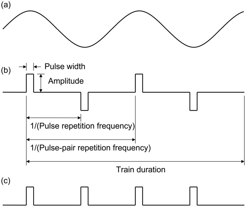
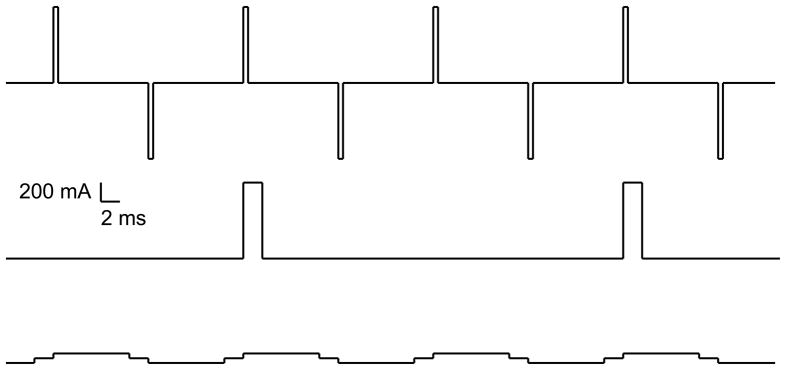
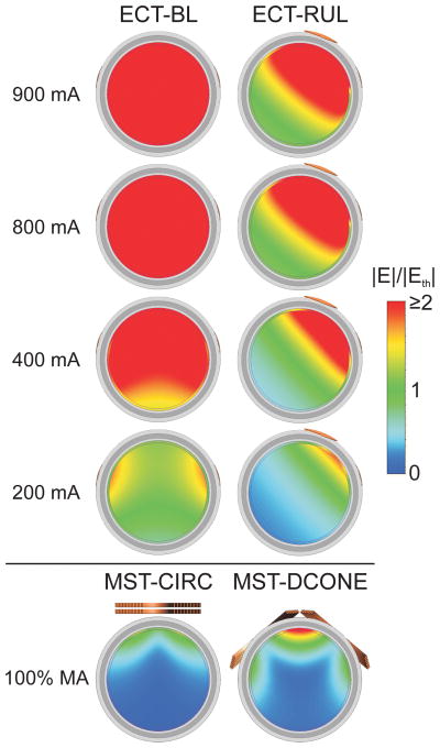
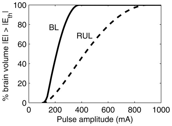
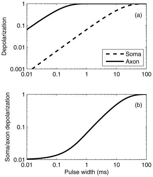
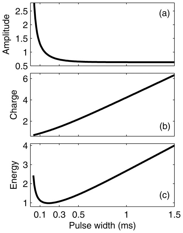
In this article, we review the parameters that define the electroconvulsive therapy (ECT) electrical stimulus and discuss their biophysical roles. We also present the summary metrics of charge and energy that are conventionally used to describe the dose of ECT and the rules commonly deployed to individualize the dose for each patient. We then highlight the limitations of these summary metrics and dosing rules in that they do not adequately capture the roles of the distinct stimulus parameters. Specifically, there is strong theoretical and empirical evidence that stimulus parameters (pulse amplitude, shape, and width, and train frequency, directionality, polarity, and duration) exert unique neurobiological effects that are important for understanding ECT outcomes. Consideration of the distinct stimulus parameters, in conjunction with electrode placement, is central to further optimization of ECT dosing paradigms to improve the risk-benefit ratio. Indeed, manipulation of specific parameters, such as reduction of pulse width and increase in number of pulses, has already resulted in dramatic reduction of adverse effects, while maintaining efficacy. Furthermore, the manipulation of other parameters, such as current amplitude, which are commonly held at fixed, high values, might be productively examined as additional means of targeting and individualizing the stimulus, potentially reducing adverse effects. We recommend that ECT dose be defined using all stimulus parameters rather than a summary metric. All stimulus parameters should be noted in treatment records and published reports. To enable research on optimization of dosing paradigms, we suggest that ECT devices provide capabilities to adjust and display all stimulus parameters.


Figures

Figure 1
Figure 1
Example ECT stimulus waveforms: (a) sine wave, (b) bidirectional rectangular pulses with indicated parameter definitions, and (c) undirectional rectangular pulses.
Figure 2
Figure 2
Three ECT stimulus trains that each have a total charge of 3.2 mC but have widely different parameters. From top down: (1) bidirectional pulse train, amplitde = 800 mA, PW = 0.5 ms, frequency = 100 pps (50 pulse-pairs per second), 8 pulses; (2) unidirectional pulse train, amplitde = 800 mA, PW = 2 ms, frequency = 25 pps, 2 pulses; (3) bidirectional pulse train, amplitde = 50 mA, PW = 8 ms, frequency = 100 pps (50 pulse-pairs per second), 8 pulses.
Figure 3
Figure 3
(a) Computer simulation with a spherical head model illustrating the electric field strength (E) relative to neural activation threshold (Eth) for conventional bilateral (BL) and right unilateral (RUL) ECT with a range of current amplitudes (PW = 0.5 ms). Coronal view is shown with tissue layers consisting of (from outer shell inward) scalp, skull, cerebrospinal fluid, gray matter, and white matter. Simulations of MST with circular (CIRC) and double-cone (DCONE) coils at 100% maximum amplitude (MA) of the Magstim Theta device (Magstim Co., Whitland, UK) are shown for comparison. Simulation methods are described by Deng et al. (b) Percentage suprathreshold stimulated brain volume as a function of ECT current amplitude derived from the spherical head model simulation.
Figure 3
Figure 3
(a) Computer simulation with a spherical head model illustrating the electric field strength (E) relative to neural activation threshold (Eth) for conventional bilateral (BL) and right unilateral (RUL) ECT with a range of current amplitudes (PW = 0.5 ms). Coronal view is shown with tissue layers consisting of (from outer shell inward) scalp, skull, cerebrospinal fluid, gray matter, and white matter. Simulations of MST with circular (CIRC) and double-cone (DCONE) coils at 100% maximum amplitude (MA) of the Magstim Theta device (Magstim Co., Whitland, UK) are shown for comparison. Simulation methods are described by Deng et al. (b) Percentage suprathreshold stimulated brain volume as a function of ECT current amplitude derived from the spherical head model simulation.
Figure 4
Figure 4
(a) Soma and axon depolarization induced by a single rectangular electric field pulse. Curves are normalized to depolarization by direct current (infinite PW). The soma and axon are assumed to have chronaxies of 10 ms and 0.1 ms, respectively. (b) Ratio of soma depolarization to axon depolarization.
Figure 5
Figure 5
Strength–duration (a), charge–duration (b), and energy–duration (c) curves for ECT, assuming neural chronaxie of 0.1 ms. The curves are normalized to unity for PW equal to the chronaxie.

References

    1. Kalinowsky LB, Hippius H. Pharmacological, convulsive, and other somatic treatments in psychiatry. New York: Grune & Stratton; 1969.
    1. Kalinowsky LB. ECT instrumentation. Biol Psychiatry. 1988;24:361–362. - PubMed
    1. Weiner RD, Rogers HJ, Davidson JR, et al. Effects of stimulus parameters on cognitive side effects. Ann N Y Acad Sci. 1986;462:315–325. - PubMed
    1. Sackeim HA, Prudic J, Devanand DP, et al. A prospective, randomized, double-blind comparison of bilateral and right unilateral electroconvulsive therapy at different stimulus intensities. Arch Gen Psychiatry. 2000;57:425–434. - PubMed
    1. Sackeim HA, Prudic J, Nobler MS, et al. Effects of pulse width and electrode placement on the efficacy and cognitive effects of electroconvulsive therapy. Brain Stimulat. 2008;1:71–83. - PMC - PubMed

Publication types