. 2010 Aug 18;5(8):e12245.
doi: 10.1371/journal.pone.0012245.

Structural insights into the quinolone resistance mechanism of Mycobacterium tuberculosis DNA gyrase

Affiliations

Structural insights into the quinolone resistance mechanism of Mycobacterium tuberculosis DNA gyrase

Jérémie Piton et al. PLoS One. .

Abstract

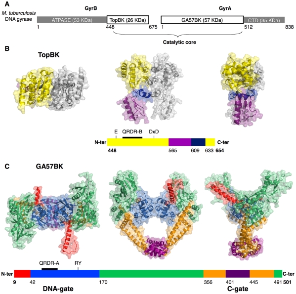
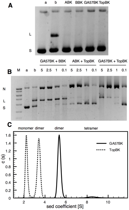
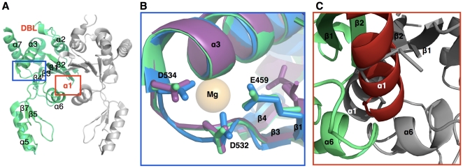
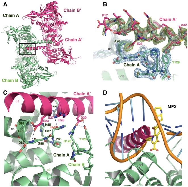
Mycobacterium tuberculosis DNA gyrase, an indispensable nanomachine involved in the regulation of DNA topology, is the only type II topoisomerase present in this organism and is hence the sole target for quinolone action, a crucial drug active against multidrug-resistant tuberculosis. To understand at an atomic level the quinolone resistance mechanism, which emerges in extensively drug resistant tuberculosis, we performed combined functional, biophysical and structural studies of the two individual domains constituting the catalytic DNA gyrase reaction core, namely the Toprim and the breakage-reunion domains. This allowed us to produce a model of the catalytic reaction core in complex with DNA and a quinolone molecule, identifying original mechanistic properties of quinolone binding and clarifying the relationships between amino acid mutations and resistance phenotype of M. tuberculosis DNA gyrase. These results are compatible with our previous studies on quinolone resistance. Interestingly, the structure of the entire breakage-reunion domain revealed a new interaction, in which the Quinolone-Binding Pocket (QBP) is blocked by the N-terminal helix of a symmetry-related molecule. This interaction provides useful starting points for designing peptide based inhibitors that target DNA gyrase to prevent its binding to DNA.


Conflict of interest statement

Competing Interests: The authors have declared that no competing interests exist.

Figures

Figure 1
Figure 1. Domain organization and structures of the individual domains from the M. tuberculosis DNA gyrase catalytic core.
A. Domain organization of the M. tuberculosis DNA gyrase. The catalytic core is composed by the Toprim domain and the breakage-reunion domain. B. Three orthogonal views of the dimeric Toprim domain from M. tuberculosis colored by regions. The crystal structure of the complete Toprim domain (TopBK) encompasses residues T448 to E654. The schematically represented primary sequence is colored as in the structure. The N-terminal residue numbers of the regions (Toprim, tail and hinge) and the TopBK C-terminal residue number are indicated. The Toprim region, constituted by discontinuous N- and C-terminal sequence segments and containing the magnesium-binding site (E459, D532 and D534) and the QRDR-B (Quinolone Resistance Determining Region in GyrB) is colored in yellow, the Tail region in purple and the hinge between the two regions in blue. The second monomer generated by a crystallographic two-fold axis is represented in grey. C. Three views of the dimeric breakage-reunion domain from M. tuberculosis colored by regions. The crystal structure of the complete breakage-reunion domain (GA57BK) extends from D9 to A501. The N-terminal helix is colored in red, the DNA-gate containing the catalytic residues R128 and Y129 and the QRDR-A in blue, the ‘tower’ in green, the helix-bundle in orange and the C-gate in purple.
Figure 2
Figure 2. Activity assays and oligomerization of the TopBK and GA57BK domains.
A. The quinolone-mediated DNA cleavage activity test measured on supercoiled pBR322 DNA (0.4 µg) as a substrate in the presence of moxifloxacin (50 µg/ml) and 2.5 µg of each subunit alone: full length subunit A (ABK), full length subunit B (BBK), GA57BK and TopBK. Lanes a and b are supercoiled pBR322 DNA and control of cleavage activity with WT M. tuberculosis DNA gyrase (ABK and BBK), respectively. B. The quinolone-mediated DNA cleavage activity test measured on supercoiled pBR322 DNA (0.4 µg) as a substrate in the presence of moxifloxacin (50 µg/ml) with various amounts (indicated by values in µg) of GA57BK associated with the full length subunit B (BBK, 1 µg), various amounts of TopBK with the full length subunit A (ABK, 1 µg), and various amounts (indicated by values in µg) of the binary complex constituted by GA57BK and TopBK. Lanes M, a and b are DNA size markers, supercoiled pBR322 DNA and control of cleavage activity with WT M. tuberculosis DNA gyrase (ABK and BBK), respectively. N, L and S denote nicked, linear and supercoiled DNA, respectively. C. Sedimentation experiments of GA57BK and TopBK. The single peak of GA57BK corresponds to the dimer, with a sedimentation coefficient of 5.4±0.2 S. The two peaks observed for TopBK correspond to the monomeric and dimeric form, with sedimentation coefficients of 2.3±0.1 S and 3.4±0.2 S, respectively. c(s) on the y-axis designates the distribution of the sedimentation coefficients observed for the experiment.
Figure 3
Figure 3. The TopBK magnesium-binding site.
A. Overall view of the dimeric structure of the Toprim domain from M. tuberculosis. One monomer constituting the asymmetric unit is represented in green, the second monomer generated by a crystallographic two-fold axis in grey. The secondary structures are indicated by black labels. The locations of the two disordered regions, the DNA Binding Loop (DBL) and the α1-helix, are indicated by the red labels “DBL” and “α1”, respectively. B. The magnesium-binding site of both M. tuberculosis TopBK structures, TopBK crystal I (3IFZ, in green) and TopBK crystal II (3M4I, in purple) with the conserved residues, E459, D532 and D534. The active site of the S. cerevisiae Toprim domain (2RGR) is represented in blue and its bound magnesium ion in orange. C. Close view of the TopBK dimer interface. The two symmetry-related α1 helices (shown in red and grey) generate steric clashes.
Figure 4
Figure 4. The active site of M. tuberculosis DNA gyrase is blocked by the N-terminal helix of a symmetry-related molecule.
A. Two dimers of GA57BK, related by the crystallographic two-fold axis, interact through the N-terminal helix. B. Omit maps for the N-terminal helix. The (2Fobs – Fcalc) map shown in blue is contoured at 1.5 σ whilst the (Fobs – Fcalc) map shown in green is contoured at 3 σ. C. Detailed interactions of the N-terminal helix (chain A', in hot pink) in the active site of the symmetry-related molecule (chain A, in light green). Y31 of the N-terminal helix and R54 of the symmetry-related molecule are located on the back-side of the helix and are not represented for better clarity. D. Based on the model discussed in the text, the N-terminal helix (chain A', in hot pink) occupies the quinolone-binding pocket (QBP) and clashes with the modeled DNA, represented in orange, and the fluoroquinolone, in yellow, bound to the QBP.
Figure 5
Figure 5. Comparison of the M. tuberculosis breakage-reunion domain to other type II topoisomerase structures.
A. Global view of the breakage-reunion domain. The boxes indicate the three close-up views shown in B, C and D. B. The DPP loop of GA57BK represented in light green is near the DNA phosphate backbone, in orange (see text for details of the model). C. Close view of the α10–α10' loop. Both M. tuberculosis structures, GA57BK (represented in light green) and MtGyrA59 (in yellow) possess a DEEX sequence insertion in this loop. The conformation of this loop is different in other bacterial type II topoisomerases, namely the three topoisomerase IV structures represented in red and E. coli GyrA59 in green, and in the three yeast topoisomerase II structures in blue. D. Close-up view of the α3–α4 loop. The conformations of GA57BK chain B (light green), and MtGyrA59 (yellow) are different from the conformation of GA57BK chain A (light green) and E. coli GyrA59 (dark green).
Figure 6
Figure 6. Model of the catalytic reaction core in complex with DNA and moxifloxacin.
A. Overall structure of the complex. GA57BK is represented in blue, TopBK in red, the DNA in orange and the moxifloxacin in green. B. Close-up view of the two quinolone-binding pockets (QBP). The purple arrow highlights the rise of the intercalated base step that constitutes the DNA walls of the QBP. Protein residues that constitute the QBP protein walls are indicated in red for TopBk and blue for GA57BK. The residues shown in sticks belong to the QRDR and are implicated in quinolone resistance. C. Close-up view along the DNA axis of one of the two QBP. The same residues as in B are represented in sticks. D. Schematic representation of the interactions between QBP residues and chemical groups of the quinolone.
Figure 7
Figure 7. Two views of the Quinolone-Binding Pocket (QBP).
The DNA-protein complex is represented in molecular surface and moxifloxacine in sticks. GA57BK is colored in dark blue, TopBK in firebrick, DNA in orange and moxifloxacin in green. The residues of TopBK belonging to the QBP are colored in yellow for the β1-α1 loop residues, in purple for the β2-DBL residues (including R482), and in pink for the DBL-α2 residues. The residues of GA57BK belonging to the QBP are represented in light green and correspond to the α3–α4 region.

References

    1. Champoux JJ. DNA topoisomerases: structure, function, and mechanism. Annu Rev Biochem. 2001;70:369–413. - PubMed
    1. Buhler C, Gadelle D, Forterre P, Wang JC, Bergerat A. Reconstitution of DNA topoisomerase VI of the thermophilic archaeon Sulfolobus shibatae from subunits separately overexpressed in Escherichia coli. Nucleic Acids Res. 1998;26:5157–5162. - PMC - PubMed
    1. Gadelle D, Filee J, Buhler C, Forterre P. Phylogenomics of type II DNA topoisomerases. Bioessays. 2003;25:232–242. - PubMed
    1. Schoeffler AJ, Berger JM. Recent advances in understanding structure-function relationships in the type II topoisomerase mechanism. Biochem Soc Trans. 2005;33:1465–1470. - PubMed
    1. Levine C, Hiasa H, Marians KJ. DNA gyrase and topoisomerase IV: biochemical activities, physiological roles during chromosome replication, and drug sensitivities. Biochim Biophys Acta. 1998;1400:29–43. - PubMed

Publication types