

The protease inhibitor alpha-2-macroglobulin-like-1 is the p170 antigen recognized by paraneoplastic pemphigus autoantibodies in human
- PMID: 20805888
- PMCID: PMC2923615
- DOI: 10.1371/journal.pone.0012250
The protease inhibitor alpha-2-macroglobulin-like-1 is the p170 antigen recognized by paraneoplastic pemphigus autoantibodies in human
Abstract
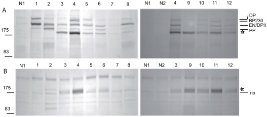
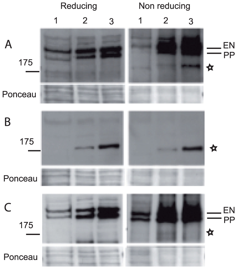
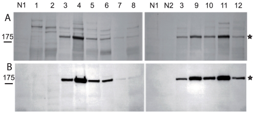
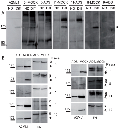
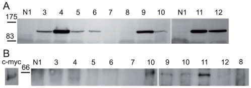
Background: Paraneoplastic pemphigus (PNP) is a devastating autoimmune blistering disease, involving mucocutaneous and internal organs, and associated with underlying neoplasms. PNP is characterized by the production of autoantibodies targeting proteins of the plakin and cadherin families involved in maintenance of cell architecture and tissue cohesion. Nevertheless, the identity of an antigen of Mr 170,000 (p170), thought to be critical in PNP pathogenesis, has remained unknown.
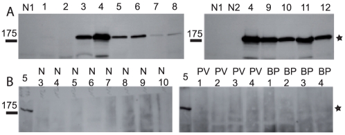
Methodology/principal findings: Using an immunoprecipitation and mass spectrometry based approach, we identified p170 as alpha-2-macroglobuline-like-1, a broad range protease inhibitor expressed in stratified epithelia and other tissues damaged in the PNP disease course. We demonstrate that 10 PNP sera recognize alpha-2-macroglobuline-like-1 (A2ML1), while none of the control sera obtained from patients with bullous pemphigoid, pemphigus vulgaris, pemphigus foliaceus and normal subjects does.
Conclusions/significance: Our study unravels a broad range protease inhibitor as a new class of target antigens in a paraneoplastic autoimmune multiorgan syndrome and opens a new challenging investigation avenue for a better understanding of PNP pathogenesis.
Conflict of interest statement
Figures
Similar articles
-
Detection of autoantibodies against alpha-2-macroglobulin-like 1 in paraneoplastic pemphigus sera utilizing novel green fluorescent protein-based immunoassays.J Dermatol Sci. 2020 Jun;98(3):173-178. doi: 10.1016/j.jdermsci.2020.04.005. Epub 2020 May 7. J Dermatol Sci. 2020. PMID: 32439251
-
Anti-α-2-macroglobulin-like-1 autoantibodies are detected frequently and may be pathogenic in paraneoplastic pemphigus.J Invest Dermatol. 2013 Jul;133(7):1785-93. doi: 10.1038/jid.2013.65. Epub 2013 Feb 13. J Invest Dermatol. 2013. PMID: 23407400
-
Human autoantibodies against HD1/plectin in paraneoplastic pemphigus.J Invest Dermatol. 1999 Feb;112(2):153-6. doi: 10.1046/j.1523-1747.1999.00498.x. J Invest Dermatol. 1999. PMID: 9989789
-
Paraneoplastic pemphigus.Australas J Dermatol. 2013 Nov;54(4):241-50. doi: 10.1111/j.1440-0960.2012.00921.x. Epub 2012 Jul 3. Australas J Dermatol. 2013. PMID: 22759072 Review.
-
[Autoimmune bullous skin diseases].Rev Med Interne. 1999 Jan;20(1):26-38. doi: 10.1016/s0248-8663(99)83006-9. Rev Med Interne. 1999. PMID: 10220817 Review. French.
Cited by
-
Paraneoplastic pemphigus: a clinical, laboratorial, and therapeutic overview.An Bras Dermatol. 2019 Oct 17;94(4):388-398. doi: 10.1590/abd1806-4841.20199165. eCollection 2019. An Bras Dermatol. 2019. PMID: 31644609 Free PMC article.
-
Significance of anti-desmocollin autoantibodies in pemphigus.J Dermatol. 2023 Feb;50(2):132-139. doi: 10.1111/1346-8138.16660. Epub 2022 Dec 28. J Dermatol. 2023. PMID: 36578135 Free PMC article. Review.
-
Molecular diagnosis in autoimmune skin blistering conditions.Curr Mol Med. 2014 Jan;14(1):69-95. doi: 10.2174/15665240113136660079. Curr Mol Med. 2014. PMID: 24160488 Free PMC article. Review.
-
A gene-centric approach to biomarker discovery identifies transglutaminase 1 as an epidermal autoantigen.Proc Natl Acad Sci U S A. 2021 Dec 21;118(51):e2100687118. doi: 10.1073/pnas.2100687118. Proc Natl Acad Sci U S A. 2021. PMID: 34911754 Free PMC article.
-
Humoral Epitope Spreading in Autoimmune Bullous Diseases.Front Immunol. 2018 Apr 17;9:779. doi: 10.3389/fimmu.2018.00779. eCollection 2018. Front Immunol. 2018. PMID: 29719538 Free PMC article. Review.
References
-
- Anhalt GJ, Kim SC, Stanley JR, Korman NJ, Jabs DA, et al. Paraneoplastic pemphigus. An autoimmune mucocutaneous disease associated with neoplasia. N Engl J Med. 1990;323:1729–1735. - PubMed
-
- Anhalt GJ. Paraneoplastic pemphigus. Adv Dermatol. 1997;12:77–96; discussion 97. - PubMed
-
- Billet SE, Grando SA, Pittelkow MR. Paraneoplastic autoimmune multiorgan syndrome: review of the literature and support for a cytotoxic role in pathogenesis. Autoimmunity. 2006;39:617–630. - PubMed
-
- Zhu X, Zhang B. Paraneoplastic pemphigus. J Dermatol. 2007;34:503–511. - PubMed
-
- Sehgal VN, Srivastava G. Paraneoplastic pemphigus/paraneoplastic autoimmune multiorgan syndrome. Int J Dermatol. 2009;48:162–169. - PubMed
Publication types
MeSH terms
Substances
LinkOut - more resources
Full Text Sources
Other Literature Sources
Medical
Molecular Biology Databases
Miscellaneous
