. 2010 Aug 27;5(8):e12441.
doi: 10.1371/journal.pone.0012441.

Autonomous bioluminescent expression of the bacterial luciferase gene cassette (lux) in a mammalian cell line

Affiliations

Autonomous bioluminescent expression of the bacterial luciferase gene cassette (lux) in a mammalian cell line

Dan M Close et al. PLoS One. .

Abstract

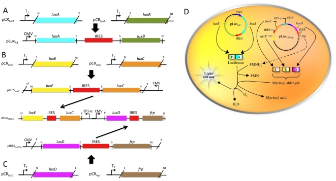
Background: The bacterial luciferase (lux) gene cassette consists of five genes (luxCDABE) whose protein products synergistically generate bioluminescent light signals exclusive of supplementary substrate additions or exogenous manipulations. Historically expressible only in prokaryotes, the lux operon was re-synthesized through a process of multi-bicistronic, codon-optimization to demonstrate for the first time self-directed bioluminescence emission in a mammalian HEK293 cell line in vitro and in vivo.

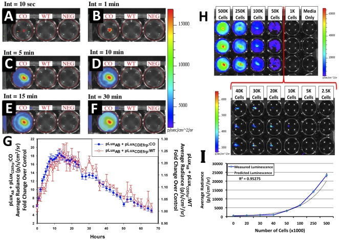
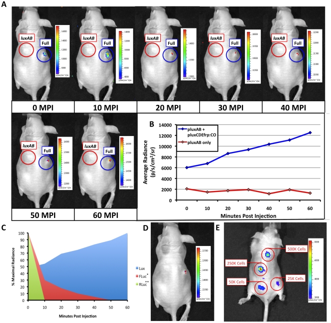
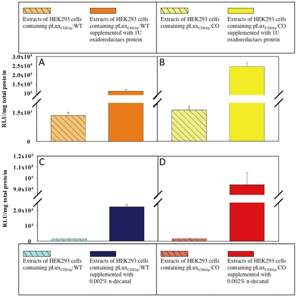
Methodology/principal findings: Autonomous in vitro light production was shown to be 12-fold greater than the observable background associated with untransfected control cells. The availability of reduced riboflavin phosphate (FMNH(2)) was identified as the limiting bioluminescence substrate in the mammalian cell environment even after the addition of a constitutively expressed flavin reductase gene (frp) from Vibrio harveyi. FMNH(2) supplementation led to a 151-fold increase in bioluminescence in cells expressing mammalian codon-optimized luxCDE and frp genes. When injected subcutaneously into nude mice, in vivo optical imaging permitted near instantaneous light detection that persisted independently for the 60 min length of the assay with negligible background.

Conclusions/significance: The speed, longevity, and self-sufficiency of lux expression in the mammalian cellular environment provides a viable and powerful alternative for real-time target visualization not currently offered by existing bioluminescent and fluorescent imaging technologies.


Conflict of interest statement

Competing Interests: The authors declare that there is no commercial conflict of interest with regards to their publication. However, it should be noted that patents #7,371,538, #7,300,792, #7,208,286, #7,090,992, #6,905,834, #6,673,596, and #6,544,729 have been awarded for which G.S.S., S.S.P., S.A.R. retain royalty rights in the event of commercialization.

Figures

Figure 1
Figure 1. Schematic showing construction and expression of the full lux cassette using a two-plasmid system.
The two-plasmid system takes advantage of IRES-based bicistronic expression to drive transcription/translation of all the genes required for autonomous bioluminescent production. (A) The pLuxAB plasmid contains the genes responsible for production of the luciferase protein. Individual luxA and luxB genes were removed from their respective vectors and ligated into the pIRES vector using the unique NheI (N) and EcoRI (E) or SalI (S) and NotI (Nt) restriction sites. (B) The pLuxCDEfrp plasmid expresses the genes required for production and regeneration of the aldehyde and FMNH2 substrates. The individual luxE and luxC genes were cloned into a pIRES vector using the unique NotI (Nt) and SalI (S) or NheI (N) and EcoRI (E) restriction sites. (C) A second pIRES vector was created that contained the luxD and frp genes inserted at the same sites. The entire luxC-IRES-luxE fragment was then inserted under the control of the EF1-α promoter in pBudCE4.1 using the unique XhoI (X) and SfiI (Sf) restriction sites, while the luxD-IRES-frp fragment was inserted under the control of the CMV promoter using the unique PstI (P) and BamHI (B) restriction sites. (D) When expressed simultaneously, these two plasmids produce all the proteins required for bioluminescence expression by utilizing the FMN and molecular oxygen stores supplied endogenously by the host cell.
Figure 2
Figure 2. in vitro bioluminescent imaging of lux cassette containing cells.
pLuxCDEfrp:CO/pLuxAB containing (CO), pLuxCDEfrp:WT/pLuxAB containing (WT), and untransfected negative control (NEG) HEK293 cells were plated in 24-well tissue culture plates and integrated for (A) 10 sec, (B) 1 min, (C) 5 min, (D) 10 min, (E) 15 min, and (F) 30 min. Bioluminescence from cells co-transfected with pLuxCDEfrp:CO/pLuxAB was distinguishable from background in the presence of untransfected cells after 10 sec and showed no increase in background detection even after a 30 min integration time. Long term in vitro expression (G) demonstrates the temporal longevity of the signal without exogenous amendment. The minimum detectable number of bioluminescent cells was determined (H) by plating a range of cell concentrations in equal volumes of media in triplicate (downward columns) in an opaque 24-well tissue culture plate. The minimum number of cells that could be consistently detected was approximately 20,000. Average radiance was shown to correlate with plated cell numbers (I), yielding an R2 value of 0.95275.
Figure 3
Figure 3. in vivo bioluminescent imaging using HEK293 cell expression of mammalian-adapted lux.
(A) HEK293 cells containing the mammalian adapted pLuxCDEfrp:CO/pLuxAB cassette (Full) were subcutaneously injected into nude mice and imaged. Detection occurred nearly immediately (<10 sec) post-injection and remained visible up to the 60 min time point of the imaging assay. HEK293 cells containing only the pLuxAB plasmid (luxAB) were subcutaneously injected into the same mouse as a negative control. Note that the automatic scaling of signal intensity differs among images, therefore creating the false appearance that image intensity is decreasing after the 10 min post-injection time point when in fact it continually increases as shown in panel (B). (C) Comparison of mammalian-adapted lux-based bioluminescence from HEK293 cells versus published data on the expression of FLuc (*[25]) and RLuc (**[8]) tagged cells over the 60 min course of the assay. (D) Upon termination of the assay 60 min post injection, the bioluminescent signal from HEK293 cells expressing the full complement of lux genes was detectable using an integration time as low as 30 sec. (E) Subcutaneous injection of HEK293 cells containing pLuxCDEfrp:CO/pLuxAB at concentrations ranging from 500,000 to 25,000 cells in 100 µl volumes of PBS demonstrated a tested lower limit of detection of 25,000 cells using a 10 min integration time. MPI, minutes post injection.
Figure 4
Figure 4. Supplementation assays demonstrating the functionality of the luxCDEfrp genes in the mammalian cell environment.
Supplementation with 1 U oxidoreductase protein significantly increased light output in cell extracts from (A) wild-type and (B) codon-optimized cell lines. Supplementation with 0.002% n-decanal resulted in increased bioluminescent output in both the (C) wild-type and (D) codon-optimized cell extracts as well, but at a lower magnitude than oxidoreductase supplementation. Values are the average of four trials, and are reported with the standard error of the mean.

References

    1. Contag CH, Bachmann MH. Advances in in vivo bioluminescence imaging of gene expression. Annu Rev Biomed Eng. 2002;4:235–260. - PubMed
    1. Oshiro M. Cooled CCD versus intensified cameras for low-light video - Applications and relative advantages. Methods in Cell Biology. 1998;56:45–62. - PubMed
    1. Tsien RY. The green fluorescent protein. Annu Rev Biochem. 1998;67:509–544. - PubMed
    1. Kalchenko V, Shivtiel S, Malina V, Lapid K, Haramati S, et al. Use of lipophilic near-infrared dye in whole-body optical imaging of hematopoietic cell homing. J Biomed Opt. 2006;11:Article #050507. - PubMed
    1. Bloch S, Lesage F, McIntosh L, Gandjbakhche A, Liang KX, et al. Whole-body fluorescence lifetime imaging of a tumor-targeted near-infrared molecular probe in mice. J Biomed Opt. 2005;10:Article #054003. - PubMed

Publication types