

Assessment of heat shock protein (HSP60, HSP72, HSP90, and HSC70) expression in cultured limbal stem cells following air lifting
- PMID: 20806039
- PMCID: PMC2927380
Assessment of heat shock protein (HSP60, HSP72, HSP90, and HSC70) expression in cultured limbal stem cells following air lifting
Abstract
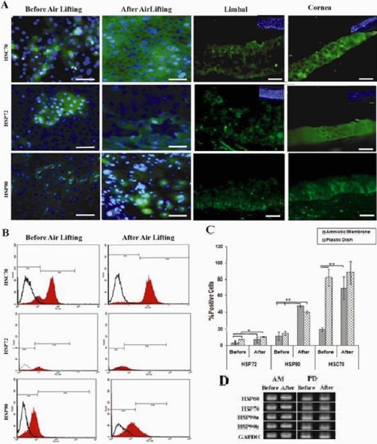
Objectives: The aim of this study is to create an ex vivo model to examine the expression of major heat-shock protein (HSP) families; HSP60, HSP72, and HSP90, and heat-shock cognate 70 (HCS70) at the mRNA and protein level in differentiating corneal cells from limbal stem cells (LSC) following air exposure.
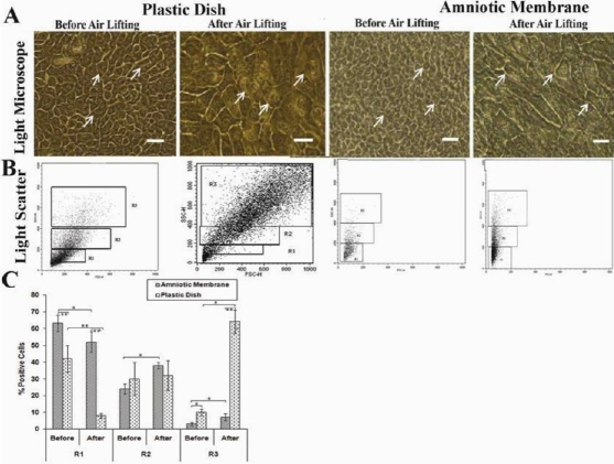
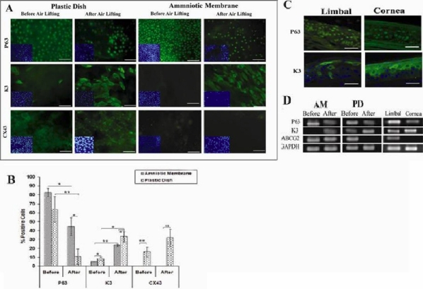
Methods: Limbal biopsies taken from cadaveric normal human limbus were cultivated as explants on human amniotic membrane (HAM) and plastic dish (PD). Corneal differentiation was induced by air lifting for 16 days. The expression of putative LSC markers (P63 and ATP-binding cassette G2 [ABCG2]), corneal markers (keratin 3 [K3/12] and connexin 43 [CX43]), and HSP60, HSP72, HSP90, and HSC70 were tested by RT-PCR, immunofluorescence, and flow cytometry pre- and post-air exposure. Fresh limbal and corneal tissues were used as control groups.
Results: Air lifting induced corneal differentiation with a decrease in the number of P63(+) cells and an increase in the number of K3(+)/CX43(+) cells, which characterized transient amplifying cells (TACs). Moreover, denuded HAM provided a superior niche for LSC proliferation and phenotype maintenance in vitro. Additionally, we have evidence that expressions of HSC70 as well as HSP72 were enhanced through corneal differentiation and HSP90 post-air lifting in vitro and in vivo. HSP60, however, was not detected in either LSC or corneal cells, in vivo and in vitro.
Conclusions: These results suggest that corneal differentiation following air exposure may regulate HSP72 and HSC70 expression. In addition, HSP72 and HSP90 may protect LSC and corneal cells against oxidative stress.
Figures
Similar articles
-
Connexin 43 expression and proliferation of human limbal epithelium on intact and denuded amniotic membrane.Invest Ophthalmol Vis Sci. 2002 Jan;43(1):63-71. Invest Ophthalmol Vis Sci. 2002. PMID: 11773014
-
Cultivation and phenotypic characterization of rabbit epithelial cells expanded ex vivo from fresh and cryopreserved limbal and oral mucosal explants.Curr Eye Res. 2015 Mar;40(3):274-81. doi: 10.3109/02713683.2014.917191. Epub 2014 May 15. Curr Eye Res. 2015. PMID: 24833207
-
Loss of corneal epithelial stem cell properties in outgrowths from human limbal explants cultured on intact amniotic membrane.Regen Med. 2008 May;3(3):329-42. doi: 10.2217/17460751.3.3.329. Regen Med. 2008. PMID: 18462056
-
Identification and characterization of limbal stem cells.Exp Eye Res. 2005 Sep;81(3):247-64. doi: 10.1016/j.exer.2005.02.016. Exp Eye Res. 2005. PMID: 16051216 Review.
-
Corneal epithelial stem cells at the limbus: looking at some old problems from a new angle.Exp Eye Res. 2004 Mar;78(3):433-46. doi: 10.1016/j.exer.2003.09.008. Exp Eye Res. 2004. PMID: 15106923 Review.
Cited by
-
Hair Follicle Generation by Injections of Adult Human Follicular Epithelial and Dermal Papilla Cells into Nude Mice.Cell J. 2017 Jul-Sep;19(2):259-268. doi: 10.22074/cellj.2016.3916. Epub 2017 Feb 22. Cell J. 2017. PMID: 28670518 Free PMC article.
-
Tissue Engineering and Regenerative Medicine in Iran: Current State of Research and Future Outlook.Mol Biotechnol. 2015 Jul;57(7):589-605. doi: 10.1007/s12033-015-9865-2. Mol Biotechnol. 2015. PMID: 25902751 Review.
-
Preventive effects of omega-3 and omega-6 Fatty acids on peroxide mediated oxidative stress responses in primary human trabecular meshwork cells.PLoS One. 2012;7(2):e31340. doi: 10.1371/journal.pone.0031340. Epub 2012 Feb 3. PLoS One. 2012. PMID: 22319624 Free PMC article.
-
Heat shock protein 72 confers protection in retinal ganglion cells and lateral geniculate nucleus neurons via blockade of the SAPK/JNK pathway in a chronic ocular-hypertensive rat model.Neural Regen Res. 2014 Jul 15;9(14):1395-401. doi: 10.4103/1673-5374.137595. Neural Regen Res. 2014. PMID: 25221598 Free PMC article.
-
The Therapeutic Roles of Recombinant Hsp90α on Cornea Epithelial Injury.Invest Ophthalmol Vis Sci. 2022 Feb 1;63(2):30. doi: 10.1167/iovs.63.2.30. Invest Ophthalmol Vis Sci. 2022. PMID: 35201262 Free PMC article.
References
-
- Walsh D, Li K, Crowther C, Marsh D, Edwards M. Thermotolerance and heat shock response during early development of the mammalian embryo. Results Probl Cell Differ. 1991;17:58–70. - PubMed
-
- Srivastava PK, Menoret A, Basu S, Binder RJ, McQuade KL. Heat shock proteins come of age: primitive functions acquire new roles in an adaptive world. Immunity. 1998;8:657–65. - PubMed
-
- Rodgers KJ, Ford JL, Brunk UT. Heat shock proteins: keys to healthy ageing? Redox Rep. 2009;14:147–53. - PubMed
-
- Christians ES, Zhou Q, Renard J, Benjamin IJ. Heat shock proteins in mammalian development. Semin Cell Dev Biol. 2003;14:283–90. - PubMed
-
- Morange M. HSFs in development. Handb Exp Pharmacol. 2006;172:153–69. - PubMed
Publication types
MeSH terms
Substances
LinkOut - more resources
Full Text Sources
Medical
Research Materials
Miscellaneous