

R213W mutation in the retinoschisis 1 gene causes X-linked juvenile retinoschisis in a large Chinese family
- PMID: 20806044
- PMCID: PMC2927431
R213W mutation in the retinoschisis 1 gene causes X-linked juvenile retinoschisis in a large Chinese family
Abstract
Purpose: We identified a large Chinese family with X-linked juvenile retinoschisis. The purpose of this study was to report the clinical findings of the family and to identify the genetic mutation by screening the retinoschisis 1 (RS1) gene.
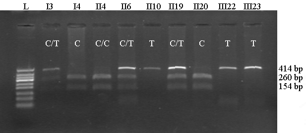
Methods: Family history was collected and all family members underwent routine ophthalmic examination. Venous blood was collected from family members and genomic DNA was extracted. The exons of RS1 were screened by PCR followed by direct sequencing and/or restriction enzyme digestion.
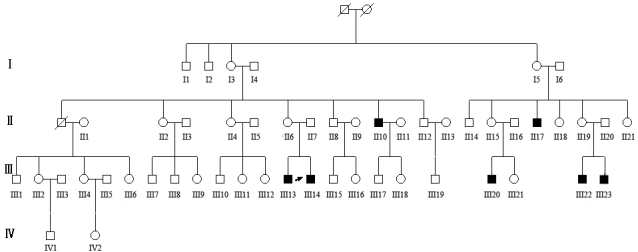
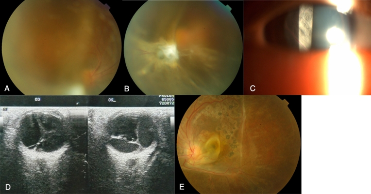
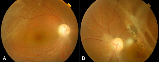
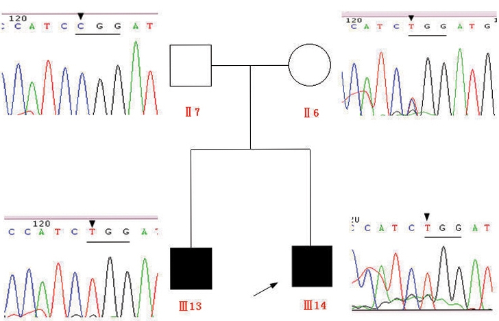
Results: The pedigree of interest was a four-generation family with 52 family members, including seven affected individuals. The proband was a 5-year-old boy showing highly elevated bullous retinoschisis with moderate vitreous hemorrhage in both eyes. Vitrectomy was performed in the left eye of the proband. Five affected males showed large peripheral retinoschisis in both eyes, either involving the macula or combined with foveal stellate cystic change. One of the affected family members showed only a foveal stellate cystic change in both eyes without periphery retinoschisis. Visual acuity of affected individuals ranged from hand motion to 0.4. The R213W mutation in exon 6 of RS1 was identified in all affected individuals, predicting an amino acid substitution of arginine to tryptophan at codon 213.
Conclusions: Our data show that the R213W mutation in RS1 causes various severities of retinoschisis in a large Chinese family, providing further evidence for X-linked juvenile retinoschisis phenotypic variability.
Figures
References
-
- The retinoschisis consortium: Functional implications of the spectrum of mutations found in 234 cases with x–linked juvenile retinoschisis. Hum Mol Genet. 1998;7:1185–92. - PubMed
-
- Wang T, Waters CT, Rothman AM, Jakins TJ, Romisch K, Trump D. Intracellular retention of mutant retinoschisin is the pathological mechanism underlying x–linked retinoschisis. Hum Mol Genet. 2002;11:3097–105. - PubMed
-
- George ND, Yates JR, Moore AT. Clinical features in affected males with x–linked retinoschisis. Arch Ophthalmol. 1996;114:274–80. - PubMed
Publication types
MeSH terms
Substances
LinkOut - more resources
Full Text Sources