

Isolation of adult progenitor cells with neuronal potential from rabbit corneal epithelial cells in serum- and feeder layer-free culture conditions
- PMID: 20806049
- PMCID: PMC2927443
Isolation of adult progenitor cells with neuronal potential from rabbit corneal epithelial cells in serum- and feeder layer-free culture conditions
Abstract
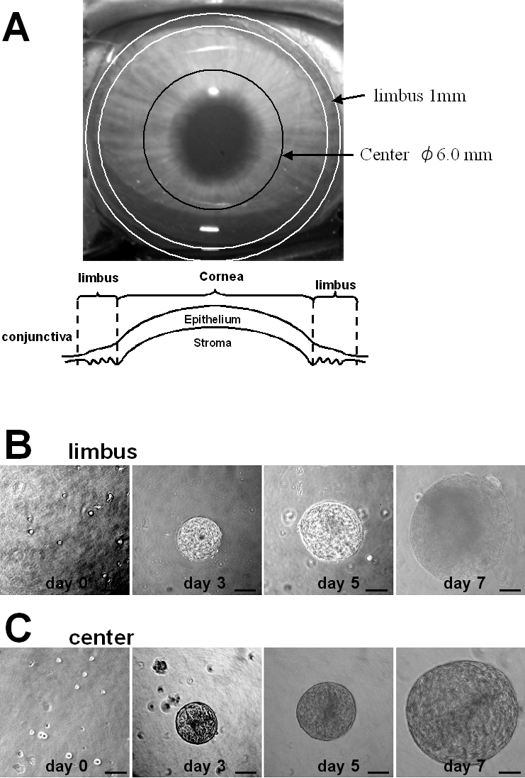
Purpose: To isolate progenitor cells from rabbit corneal epithelial cells (CEC) in serum- and feeder layer-free culture conditions and to compare the self-renewal capacity of corneal epithelial progenitor cells obtained from the central and limbal regions of the cornea.
Methods: Tissue samples of New Zealand white rabbit corneas were dissected from the limbal and central regions to obtain CEC for sphere-forming culture, in which the cells formed spheres in serum-free medium containing growth factors. The number of primary and secondary sphere colonies and the size of the primary spheres were compared between the limbal and central regions. To promote differentiation, isolated sphere colonies were plated in dishes coated with poly-L-lysine (PLL)/laminin. The expression of epithelial, neural, and mesenchymal mRNAs was examined in the sphere colonies and their progeny by immunocytochemistry and/or the reverse transcription-polymerase chain reaction (RT-PCR). Adherent differentiated cells from the sphere colonies were also examined morphologically.
Results: Primary spheres were isolated from both the limbal and central regions of the cornea. The rate of primary sphere formation by CEC from the limbal region (55.6+/-10.6/10,000 cells) was significantly higher than that by cells from the central cornea (43.1+/-7.2/10,000 cells, p=0.0028), but there was no significant difference in the size of primary spheres derived from both regions. The self-renewal capacity of cells from the limbal region was higher than that of cells from the central region, as evidenced by the significantly higher secondary sphere formation rate of limbal cells (38.7+/-8.5/10,000 cells) in comparison with that for central cells (31.3+/-5.7/10,000 cells, p=0.013). The primary sphere colonies expressed bromodeoxyuridine (BrdU), a 63-kDa protein (p63), p75 neurotrophin receptor (p75(NTR)), and nestin, whereas their progeny expressed cytokeratin 3, cytokeratin 12, vimentin, alpha-smooth muscle actin, microtubule-associated protein 2, and neuron-specific enolase on immunocytochemical analysis. These markers were confirmed by RT-PCR.
Conclusions: Our findings indicate that limbal CEC contain more progenitor cells with a stronger self-renewal capacity than cells from the central region. These progenitor cells differentiate into the epithelial lineage, and can also produce neuronal protein.
Figures


Similar articles
-
Human corneal endothelial cell precursors isolated by sphere-forming assay.Invest Ophthalmol Vis Sci. 2005 May;46(5):1626-31. doi: 10.1167/iovs.04-1263. Invest Ophthalmol Vis Sci. 2005. PMID: 15851561
-
Easy xeno-free and feeder-free method for isolating and growing limbal stromal and epithelial stem cells of the human cornea.PLoS One. 2017 Nov 17;12(11):e0188398. doi: 10.1371/journal.pone.0188398. eCollection 2017. PLoS One. 2017. PMID: 29149196 Free PMC article.
-
Isolation and distribution of rabbit keratocyte precursors.Mol Vis. 2008 Jan 30;14:197-203. Mol Vis. 2008. PMID: 18334932 Free PMC article.
-
Characterization, isolation, expansion and clinical therapy of human corneal epithelial stem/progenitor cells.J Stem Cells. 2014;9(2):79-91. J Stem Cells. 2014. PMID: 25158157 Review.
-
Isolation of human corneal endothelial cell precursors and construction of cell sheets by precursors.Cornea. 2006 Dec;25(10 Suppl 1):S90-2. doi: 10.1097/01.ico.0000247221.95424.d7. Cornea. 2006. PMID: 17001202 Review.
Cited by
-
Role of Human Corneal Stroma-Derived Mesenchymal-Like Stem Cells in Corneal Immunity and Wound Healing.Sci Rep. 2016 May 19;6:26227. doi: 10.1038/srep26227. Sci Rep. 2016. PMID: 27195722 Free PMC article.
-
Spatial distribution of niche and stem cells in ex vivo human limbal cultures.Stem Cells Transl Med. 2014 Nov;3(11):1331-41. doi: 10.5966/sctm.2014-0120. Epub 2014 Sep 17. Stem Cells Transl Med. 2014. PMID: 25232182 Free PMC article.
References
-
- Potten CS, Schofield R, Lajtha LG. A comparison of cell replacement in bone marrow, testis and three regions of surface epithelium. Biochim Biophys Acta. 1979;560:281–99. - PubMed
-
- Lavker RM, Sun TT. Heterogeneity in epidermal basal keratinocytes: morphological and functional correlations. Science. 1982;215:1239–41. - PubMed
-
- Lavker RM, Sun TT. Epidermal stem cells. J Invest Dermatol. 1983;81:121s–7s. - PubMed
-
- Ferraris C, Chevalier G, Favier B, Jahoda CA, Dhouailly D. Adult corneal epithelium basal cells possess the capacity to activate epidermal, pilosebaceous and sweat gland genetic programs in response to embryonic dermal stimuli. Development. 2000;127:5487–95. - PubMed
-
- Kinoshita S, Friend J, Thoft RA. Sex chromatin of donor corneal epithelium in rabbits. Invest Ophthalmol Vis Sci. 1981;21:434–41. - PubMed
Publication types
MeSH terms
Substances
LinkOut - more resources
Full Text Sources
Research Materials