. 2010 Jul 21;1(7):505-519.
doi: 10.1021/cn1000217.

Mapping neuropeptide expression by mass spectrometry in single dissected identified neurons from the dorsal ganglion of the nematode Ascaris suum

Affiliations

Mapping neuropeptide expression by mass spectrometry in single dissected identified neurons from the dorsal ganglion of the nematode Ascaris suum

Jessica L Jarecki et al. ACS Chem Neurosci. .

Abstract

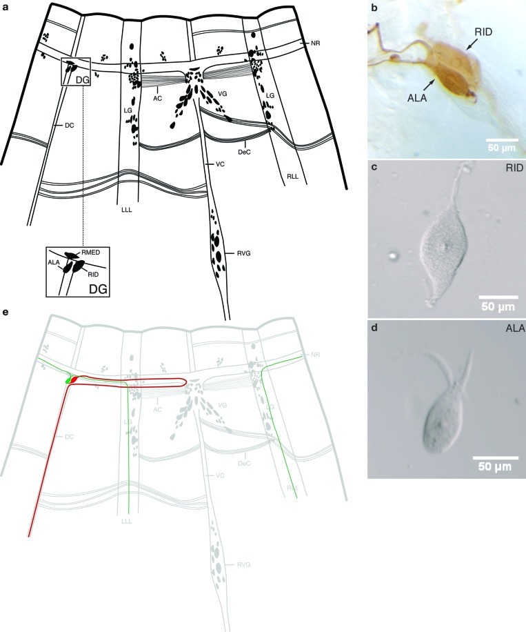
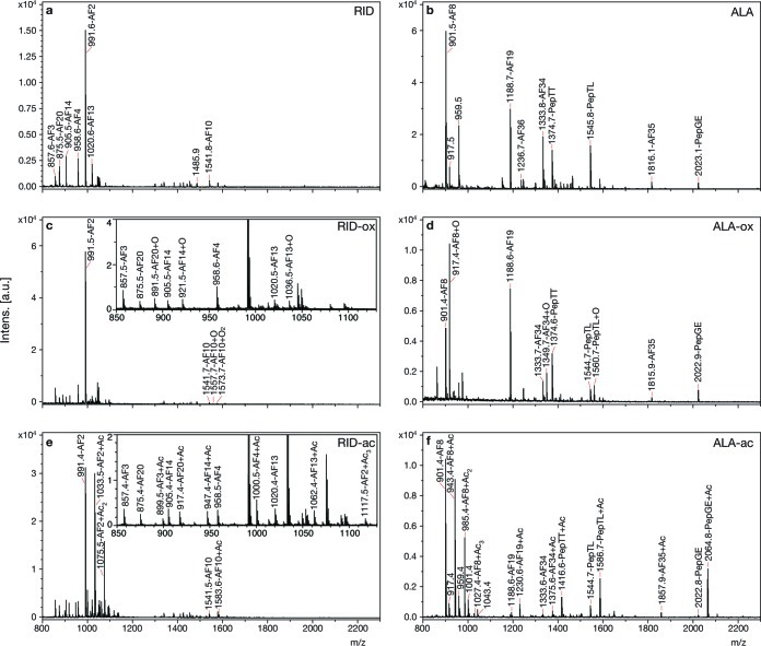
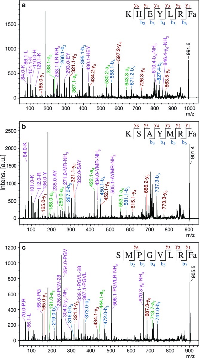
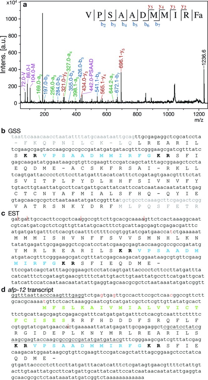
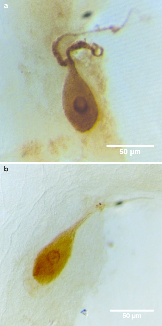
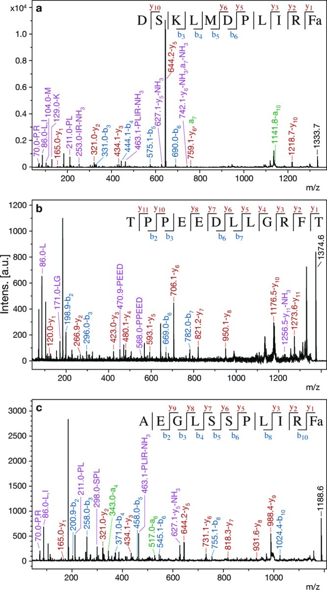
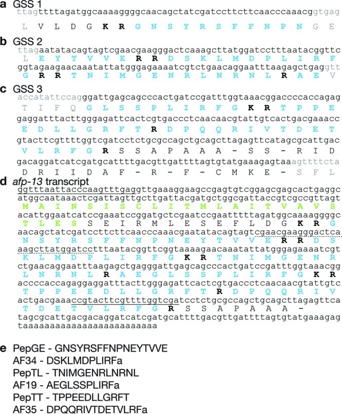
We have developed a method for dissecting single neurons from the nematode Ascaris suum, in order to determine their peptide content by mass spectrometry (MS). In this paper, we use MALDI-TOF MS and tandem MS to enumerate and sequence the peptides present in the two neurons, ALA and RID, that comprise the dorsal ganglion. We compare the peptide content determined by MS with the results of immunocytochemistry and in situ hybridization of previously isolated peptides AF2, AF8 and 6 peptides encoded by the afp-1 transcript. We find complete agreement between the three techniques, which validates single neuron MS as a method for peptide localization. We also discovered and sequenced 6 novel peptides in the ALA neuron. Cloning of cDNAs and database searching of Genomic Survey Sequences showed that transcript afp-12 encodes peptide AF36 (VPSAADMMIRFamide), and afp-13 encodes AF19 (AEGLSSPLIRFamide), AF34 (DSKLMDPLIRFamide), AF35 (DPQQRIVTDETVLRFamide), and 3 non-amidated peptides (PepTT, PepTL, and PepGE). We have found no similarities with reported peptide expression in the nematode Caenorhabditis elegans. This method promises to be ideally suited for determining the peptide content of each of the 298 neurons in the nervous system of this nematode.


Figures

Figure 1
Figure 1
The neurons of the dorsal ganglion (DG). (a) Diagram of the head ganglia of A. suum, modified from Goldschmidt (63). The worm has been split near the dorsal axis and opened flat. Neuronal cell bodies and commissural processes are shown. The nerve ring (NR), ventral ganglion (VG), dorsal ganglion (DG), lateral ganglia (LG), and retrovesicular ganglion (RVG) are indicated. LLL, left lateral line; RLL, right lateral line; VC, ventral nerve cord; DC, dorsal nerve cord; DeC, deirid commissures; AC, amphidial commissures. The inset shows higher magnification of the identified neurons ALA and RID in the DG. Neuron RMED lies within the NR, not the DG. (b) Immunological staining (ICC) of DG in a whole mount of the head region using anti-FMRFa antibody (25), which stains both cells of the DG. (c) Dissected unstained individual RID. (d) Dissected unstained individual ALA. (e) Diagram showing the cell bodies and neural processes of ALA (green) and RID (red).
Figure 2
Figure 2
Neuropeptides from single DG neurons in native and chemically modified forms. (a) Mass spectrum from an individual RID with no chemical modifications. (b) Spectrum from an individual ALA with no chemical modifications. (c) Spectrum from an individual RID stained with methylene blue, which oxidizes methionines and adds 16 mass units. The inset is an expansion of the small peaks between 850 and 1130 m/z. (d) Spectrum from an individual ALA stained with methylene blue. (e) Spectrum from an individual RID treated with acetic anhydride, which acetylates free amines (N-terminus, lysine) and to a lesser extent tyrosine and adds 42 mass units. The inset is an expansion of the small peaks between 850 and 1130 m/z. (f) Spectrum from an individual ALA treated with acetic anhydride. X axis, m/z, is mass-to-charge ratio. Y axis is intensity of MS signal in arbitrary units, a.u.
Figure 3
Figure 3
MS/MS spectra of peptides with known expression in DG. Peaks representing a (green), b (blue), y (red), and high-intensity internal fragment (purple) ions are labeled, and b and y ions are summarized in the sequence at the top of each spectrum. (a) MS/MS spectrum from RID of AF2 (afp-4), m/z 991.6. (b) MS/MS spectrum from ALA of AF8 (afp-3), m/z 901.4. (c) MS/MS spectrum from RID of AF14 (afp-1), m/z 905.5.
Figure 4
Figure 4
AF36 and afp-12. (a) MS/MS spectrum from ALA of AF36, m/z 1236.6. Peaks representing a (green), b (blue), y (red), and high-intensity internal fragment (purple) ions are labeled, and b and y ions are summarized in the sequence at the top of the spectrum. (b) Sequence from GSS database encoding AF36. (c) Sequence from EST library encoding AF36 (CB040272 (30)). (d) Cloned afp-12 transcript deduced by unique primers and 3′ RACE (HM125966). Gray indicates suspected intronic region. Blue indicates encoded peptide. Bold indicates basic cleavage sites. Green indicates signal peptide. Sequences used to design primers for PCR have a solid underline; the 5′ primer was SL1. Nucleotides with a dotted underline were used for the primer for 3′ RACE. In panels c and d red bases and vertical lines indicate insertions/deletions between the two sequences.
Figure 5
Figure 5
Confirmation by ICC of peptides localized in ALA. (a) ICC with anti-AF36 antibody. (b) ICC with anti-AF19 antibody.
Figure 6
Figure 6
MS/MS of peptides encoded by afp-13. Peaks representing a (green), b (blue), y (red), and high-intensity internal fragment (purple) ions are labeled, and b and y ions are summarized in the sequence at the top of each spectrum. (a) MS/MS spectrum from ALA of AF34, m/z 1333.7. (b) MS/MS spectrum from ALA of PepTT, m/z 1374.6. (c) MS/MS spectrum from ALA of AF19, m/z 1188.6.
Figure 7
Figure 7
Nucleotide sequence and deduced amino acid sequence of afp-13: (a) Sequence from GSS database (GSS 1) encoding a portion of PepGE. (b) GSS 2 encoding AF34, PepTL, a small portion of AF19, and the remaining portion of PepGE. (c) GSS 3 encoding PepTT, AF35, and the remaining portion of AF19. (d) Cloned afp-13 transcript deduced by unique primers and 3′ RACE (HM125967). (e) Processed peptides from afp-13. Gray indicates predicted intronic region. Blue indicates encoded peptide. Bold indicates basic cleavage sites. Green indicates signal peptide. Sequences used to design primers for PCR have a solid underline; the 5′ primer was SL1. Nucleotides with a dotted underline were used for the primer for 3′ RACE.

Similar articles

Cited by

References

    1. Kotani M.; Detheux M.; Vandenbogaerde A.; Communi D.; Vanderwinden J. M.; Le Poul E.; Brezillon S.; Tyldesley R.; Suarez-Huerta N.; Vandeput F.; Blanpain C.; Schiffmann S. N.; Vassart G.; Parmentier M. (2001) The metastasis suppressor gene KiSS-1 encodes kisspeptins, the natural ligands of the orphan G protein-coupled receptor GPR54. J. Biol. Chem. 276, 34631–34636. - PubMed
    1. Cottrell G. A. (1997) The first peptide-gated ion channel. J. Exp. Biol. 200, 2377–2386. - PubMed
    1. Marder E.; Bucher D. (2007) Understanding circuit dynamics using the stomatogastric nervous system of lobsters and crabs. Annu. Rev. Physiol. 69, 291–316. - PubMed
    1. Stretton A.; Donmoyer J.; Davis R.; Meade J.; Cowden C.; Sithigorngul P. (1992) Motor behavior and motor nervous system function in the nematode Ascaris suum. J. Parasitol. 78, 206–214. - PubMed
    1. Davis R. E.; Stretton A. O. (2001) Structure-activity relationships of 18 endogenous neuropeptides on the motor nervous system of the nematode Ascaris suum. Peptides 22, 7–23. - PubMed