. 2010 Aug 4:16:1467-74.

Resveratrol protects human lens epithelial cells against H2O2-induced oxidative stress by increasing catalase, SOD-1, and HO-1 expression

Affiliations

Resveratrol protects human lens epithelial cells against H2O2-induced oxidative stress by increasing catalase, SOD-1, and HO-1 expression

Yi Zheng et al. Mol Vis. .

Abstract

Purpose: Oxidative damage induced by H(2)O(2) treatment can irreversibly damage the lens epithelium, resulting in cell death and cataract. Whether the effects of oxidative stress could be attenuated in cultured human lens epithelial cells by incubation with resveratrol (RES) is still unknown. In the present study, we examined the function of resveratrol in protecting human lens epithelial B-3 (HLEB-3) cells against H(2)O(2) induced cell death and cell apoptosis, its role in reducing H(2)O(2) induced intracellular reactive oxygen species (ROS) accumulation, and investigated the mechanism by which resveratrol underlies the effect.

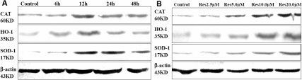
Methods: HLEB-3 cells, a human lens epithelial cell line, were exposed to 100 muM H(2)O(2) with or without RES pre-treatment at different concentrations for different time duration. Cell viabilities were monitored by 4-[3-[4-iodophenyl]-2-4(4-nitrophenyl)-2H-5-tetrazolio-1,3-benzene disulfonate] (WST-1) assay. The apoptosis rate and ROS generation were detected by flow cytometric analysis. Expression levels of superoxide dismutases-1 (SOD-1), catalase, and heme oxygenase-1 (HO-1) proteins were measured by western-blotting analysis. p38 and c-jun N terminal kinase (JNK) activation was also evaluated by western-blotting analysis.

Results: Resveratrol clearly reduced H(2)O(2) induced cell apoptosis and ROS accumulation; protected HLEB-3 cells from H(2)O(2) induced oxidative damage, and increased the expression levels of SOD-1, catalase, and HO-1. Further studies showed that RES also inhibited H(2)O(2) induced p38 and JNK phosphorylation.

Conclusions: These findings suggested that RES protected HLEB-3 cells from H(2)O(2) induced oxidative damage, presumably by inducing three antioxidative enzymes including catalase, SOD-1, and HO-1.


Figures

Figure 1
Figure 1
Resveratrol protects against H2O2-induced cell death and apoptosis in HLEB-3 cells. A: The HLEB-3 cells were incubated with different concentrations of RES (2.5, 5.0, 10.0, and 20.0 μM) for12 h before H2O2 (100 μM) treatment for 24 h. B: The cells were incubated with 20.0 μM RES for different time (12, 24, 48 h), then treated with 100 μM H2O2 for 24 h. C: The cells were pretreated with 20.0 μM RES for 12 h,then exposed to different concentration of H2O2 (50, 100, 200 μM) for 24 h, the cell viability was measured by WST-1 assay. The data are represented as means±SD from three independent experiments. The asterisk indicates that p<0.05 compared to the untreated control. D: The cells were incubated with 10.0 and 20.0 μM RES for 12 h then treated with 100 μM H2O2 for 24 h. Flow cytometric analysis was used to quantify the rate of cell apoptosis using double staining of Annexin V-FITC and PI. The result is one representative example of three separate experiments. E: Resveratrol (20μM) significantly decreased the apoptosis rate of HLEB-3 cells, **p<0.01 versus positive control (t-test), the preventative effect of 20 μM RES was improved versus 10 μM RES,*p<0.05.
Figure 2
Figure 2
Resveratrol reduced the generation of ROS. A: The cells were pretreated with 10.0 and 20.0 μΜ RES for 12 h followed by treatment with 100 μM H2O2 for 2 h. The production of ROS was examined by measuring the level of ROS production using DCF-DA by flow cytometry. The result is one representative example of three separate experiments. B: Resveratrol (20.0 μM) significantly reduced the ROS generation in HLEB-3 cells **p<0.01 versus the RES negative control. The degree of 20.0 μM RES was higher than 10.0 μM RES *p<0.05.
Figure 3
Figure 3
Resveratrol upregulated the expression of SOD-1, HO-1, and catalase. A: western blotting analysis of SOD-1, HO-1, and catalase at different time points (6, 12, 24, and 48 h) after pretreatment with 20.0 μM resveratrol. B: western blotting analysis of SOD-1, HO-1, and catalase 12 h after pretreatment with different concentration of resveratrol (2.5, 5.0, 10.0, and 20.0 μM). The expression levels of RES pretreated cells significantly increased compared with control cells (p<0.05), and the expression levels were proportional to the concentration of resveratrol.
Figure 4
Figure 4
Resveratrol inhibits p38 and JNK phosphorylation. HLEB-3 cells were treated with 100 μM H2O2 with or without 20.0 μM resveratrol for intervals ranging between 1 and 3 h and p38 and JNK activities were evaluated by western blotting analysis using anti-phosphorylated p38 and anti-phosphorylated JNK antibodies. Resveratrol significantly inhibited p38 and JNK phosphorylation compared to control. Anti-p38 and anti-JNK protein antibodies show equal loading of protein samples.

Similar articles

Cited by

References

    1. Beatty S, Koh H, Phil M, Henson D, Boulton M. The role of oxidative stress in the pathogenesis of age-related macular degeneration. Surv Ophthalmol. 2000;45:115–34. - PubMed
    1. Giblin FJ, Boyle DL, Takemoto LJ, Ho YS, Knoernschild T. Juenemann,Glutathione: a vital lens antioxidant. J Ocul Pharmacol Ther. 2000;16:121–35. - PubMed
    1. Phelps Brown N, Bron AJ. Lens disorders: a clinical manual of cataract diagnosis. Oxford: Butterworth-Heinemann; 1996.
    1. Brady JP, Garland D, Duglas-Tabor Y, Robison WG, Jr, Groome A, Wawrousek EF. Targeted disruption of the mouse alpha A-crystallin gene induces cataract and cytoplasmic inclusion bodies containing the small heat shock protein alpha B-crystallin. Proc Natl Acad Sci USA. 1997;94:884–9. - PMC - PubMed
    1. Reddy VN, Giblin FJ, Lin LR, Dang L, Unakar NJ, Musch DC. A, Lutjen-Drecoll E. Glutathione peroxidase-1 deficiency leads to increased nuclear light scattering, membrane damage, and cataract formation in gene-knockout mice. Invest Ophthalmol Vis Sci. 2001;42:3247–55. - PubMed

Publication types

MeSH terms

LinkOut - more resources