

Ultrasonographic findings in Cairn Terriers with preclinical renal dysplasia
- PMID: 20806879
- PMCID: PMC3235696
- DOI: 10.1111/j.1740-8261.2010.01674.x
Ultrasonographic findings in Cairn Terriers with preclinical renal dysplasia
Abstract
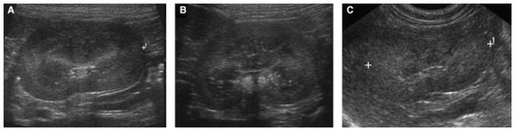
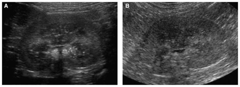
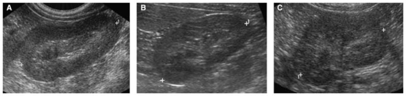
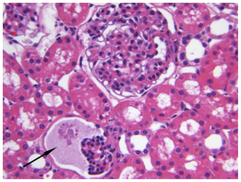
Renal dysplasia is a hereditary disease characterized by abnormal differentiation of renal tissue. The ultrasonographic appearance of dysplastic canine kidneys has been reported in the late stage of the disease where inflammatory and degenerative changes are already present and the dogs are in chronic renal failure. In this study, we describe the ultrasonographic appearance of the kidneys of five related Cairn Terriers affected with renal dysplasia before the onset of clinical or laboratory evidence of renal failure. Common findings included poor corticomedullary definition and multifocal hyperechoic speckles in the renal medulla, or a diffusely hyperechoic medulla. Severity of ultrasonographic changes was related to the severity of histopathologic findings. The ability to detect dysplastic changes before clinical signs develop makes ultrasound a potentially useful screening method for canine renal dysplasia.
Figures
Similar articles
-
Ultrasonographic findings of renal dysplasia in cocker spaniels: eight cases.Acta Vet Hung. 1997;45(4):397-408. Acta Vet Hung. 1997. PMID: 9557317
-
Ultrasonographic characteristics of canine renal lymphoma.Vet Radiol Ultrasound. 2014 Jul-Aug;55(4):441-6. doi: 10.1111/vru.12152. Epub 2014 Mar 14. Vet Radiol Ultrasound. 2014. PMID: 24629062
-
Polycystic disease of the kidney and liver in the Cairn Terrier.Vet Pathol. 1980 Jul;17(4):436-42. doi: 10.1177/030098588001700405. Vet Pathol. 1980. PMID: 7385577
-
B-mode and Doppler ultrasound of chronic kidney disease in dogs and cats.Vet Res Commun. 2017 Dec;41(4):307-315. doi: 10.1007/s11259-017-9694-9. Epub 2017 Jun 21. Vet Res Commun. 2017. PMID: 28634673 Review.
-
[A clinical approach to renal cysts].Rev Med Chil. 2007 Jan;135(1):111-20. doi: 10.4067/s0034-98872007000100016. Epub 2007 Mar 6. Rev Med Chil. 2007. PMID: 17369992 Review. Spanish.
Cited by
-
Case report: BCL-2 and CD31 immunoexpression related to clinical and histopathological evaluation of renal dysplasia in a Welsh Corgi Puppy.Front Vet Sci. 2022 Oct 4;9:995765. doi: 10.3389/fvets.2022.995765. eCollection 2022. Front Vet Sci. 2022. PMID: 36268045 Free PMC article.
-
DNA Sequence Variants in the Five Prime Untranslated Region of the Cyclooxygenase-2 Gene Are Commonly Found in Healthy Dogs and Gray Wolves.PLoS One. 2015 Aug 5;10(8):e0133127. doi: 10.1371/journal.pone.0133127. eCollection 2015. PLoS One. 2015. PMID: 26244515 Free PMC article.
References
-
- Greco DS. Congenital and inherited renal disease of small animals. Vet Clin North Am Small Anim Pract. 2001;31:393–399. - PubMed
-
- Abraham LA, Beck C, Slocombe RF. Renal dysplasia and urinary tract infection in a Bull Mastiff puppy. Aust Vet J. 2003;81:336–339. - PubMed
-
- De Morais HS, DiBartola SP, Chew DJ. Juvenile renal disease in Golden Retrievers: 12 cases (1984–1994) J Am Vet Med Assoc. 1996;209:792–797. - PubMed
-
- Felkai CS, Voros K, Vrabely T, Vetesi F, Karsai F, Papp L. Ultrasonographic findings of renal dysplasia in Cocker Spaniels: eight cases. Acta Veterinaria Hungarica. 1997;45:397–408. - PubMed
-
- Hoppe A, Karlstam E. Renal dysplasia in Boxers and Finnish Harriers. J Small Anim Pract. 2000;41:422–426. - PubMed
MeSH terms
Grants and funding
LinkOut - more resources
Full Text Sources
Medical
