. 2010 Nov;78(4):866-82.
doi: 10.1111/j.1365-2958.2010.07369.x. Epub 2010 Sep 23.

Nucleoid occlusion prevents cell division during replication fork arrest in Bacillus subtilis

Affiliations

Nucleoid occlusion prevents cell division during replication fork arrest in Bacillus subtilis

Remi Bernard et al. Mol Microbiol. 2010 Nov.

Abstract

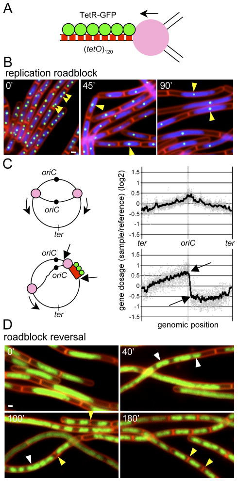
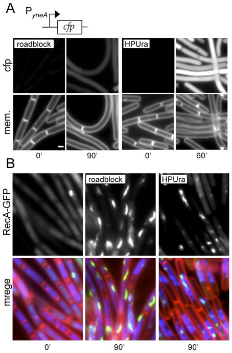
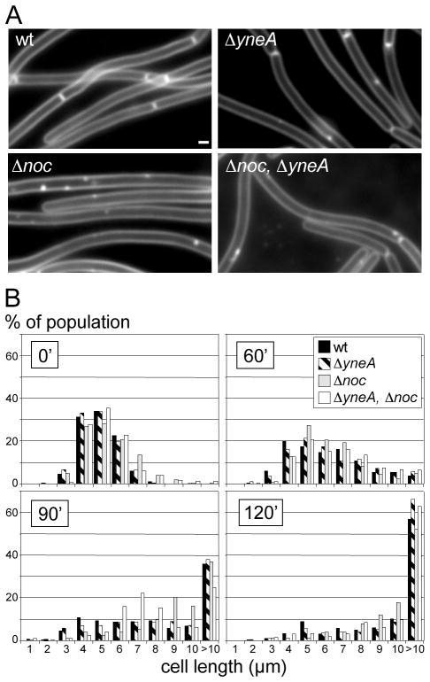
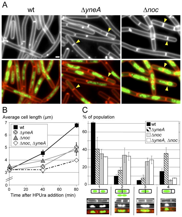
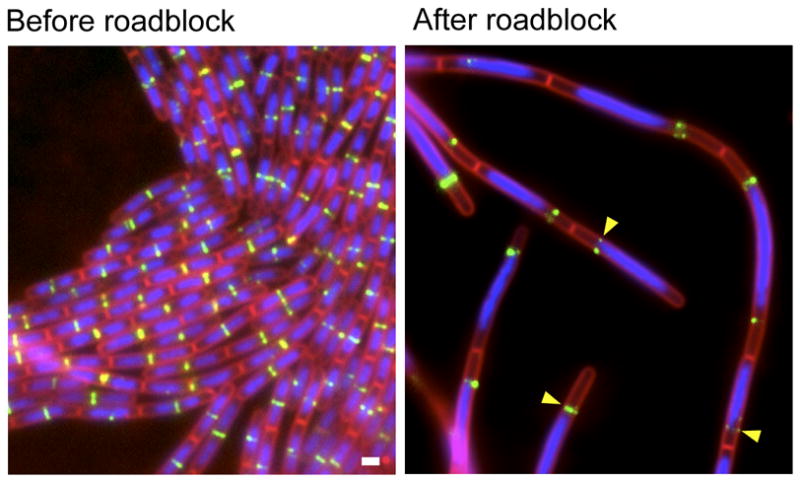
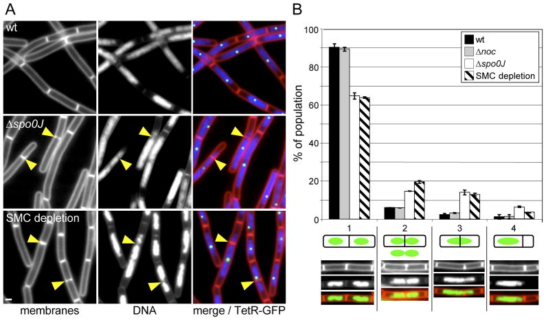
How bacteria respond to chromosome replication stress has been traditionally studied using temperature-sensitive mutants and chemical inhibitors. These methods inevitably arrest all replication and lead to induction of transcriptional responses and inhibition of cell division. Here, we used repressor proteins bound to operator arrays to generate a single stalled replication fork. These replication roadblocks impeded replisome progression on one arm, leaving replication of the other arm and re-initiation unaffected. Remarkably, despite robust generation of RecA-GFP filaments and a strong block to cell division during the roadblock, patterns of gene expression were not significantly altered. Consistent with these findings, division inhibition was not mediated by the SOS-induced regulator YneA nor by RecA-independent repression of ftsL. In support of the idea that nucleoid occlusion prevents inappropriate cell division during fork arrest, immature FtsZ-rings formed adjacent to the DNA mass but rarely on top of it. Furthermore, mild alterations in chromosome compaction resulted in cell division that guillotined the DNA. Strikingly, the nucleoid occlusion protein Noc had no discernable role in division inhibition. Our data indicate that Noc-independent nucleoid occlusion prevents inappropriate cell division during replication fork arrest. They further suggest that Bacillus subtilis normally manages replication stress rather than inducing a stress response.


Figures

Figure 1
Figure 1
TetR bound to tetO arrays causes a reversible replication block. (A) Schematic representation of a stalled replication fork generated by TetR-GFP (green circles) bound to an array of tet operators (red boxes). The repressor proteins block replisome (pink circle) progression. (B) Images before and after induction of a replication roadblock at +7° (81.7 kb from the origin of replication) in strain BRB63. Time (in min) after removal of the inducer aTC is indicated. Images show membranes stained with FM4-64 (red), DAPI-stained DNA (blue), and TetR-GFP (green) bound to (tetO)120. The reduction in TetR-GFP foci (and therefore the number of +7° loci per cell) following the removal of aTC is highlighed (yellow carets). White bar is 1 μm. (C) Genomic microarray analysis of strain BRB63 before (upper panel) and after (lower panel) the replication roadblock. Gene dosage (log2) relative to a reference DNA is on the y-axis. All the probed genes in the B. subtilis chromosome arranged from −188° to +172° (ter-oriC-ter) are shown (grey dots). The smooth line was generated by plotting the average gene dosage of the 25 genes before and 25 genes after each gene probed. Arrows indicate a position before (+6°) and after (+8°) the site of insertion of the (tetO)120 array (+7°). Schematic representations of the two conditions are shown to the left of the graphs. (D) Chromosome segregation and cell division upon release of fork arrest. The replication roadblock was induced for 90 min in strain BRB63. aTC was added to the culture to release the replication fork and cells were visualized by fluorescence microscopy at the times indicated. Images show membranes stained with FM4-64 (red) and DAPI-stained DNA (false-colored green). New cell division events (yellow carets) and resolution and segregation of the DNA mass into distinct nucleoids (white carets) are highlighted.
Figure 2
Figure 2
The replication roadblock generates RecA-GFP foci and filaments but does not significantly induce the SOS response. (A) Visualization of an SOS-reporter (PyneA-cfp) in response to a replication roadblock (left panel; strain BRB190) or following HPUra treatment (right panel; strain BRB175). Membranes (mem.) were visualized with FM4-64. Exposure times for the fluorescent reporter were identical in all strains and the fluorescent intensities were scaled identically. White bar is 1 μm. A TetR-YFP fusion was used instead of TetR-GFP to generate the roadblock to ensure the emission spectra of the SOS reporter and fluorescent repressor protein were distinct. TetR-YFP was as efficient as TetR-GFP in blocking replication fork progression and inhibiting cell division (data not shown). (B) Localization of RecA-GFP in cells before and after replication arrest in response to a replication roadblock (BRB636, middle panel) or to HPUra (BDR2429, right panel). The left panel shows exponentially growing cells from BDR2429 before addition of HPUra. A time course of RecA-GFP foci/filament formation is shown in Figure S6B. Images show membranes stained with FM4-64 (red), DAPI-stained DNA (blue) and RecA-GFP (green). A TetR-CFP fusion was used instead of TetR-GFP to generate the replication roadblock. TetR-CFP was slightly less efficient at blocking replication fork progression as TetR-GFP (data not shown).
Figure 3
Figure 3
Cell division inhibition during the replication roadblock is independent of YneA and Noc. (A) Representative images of cell filaments after 120′ of replication fork arrest in wild-type (BRB1), or strains lacking yneA (ΔyneA; BRB35), noc (Δnoc; BRB117), or both (Δnoc, ΔyneA; BRB117). White bar is 1 μm. (B) Histograms show the length distribution of the indicated strains during fork arrest. Time (in min) after induction of the roadblock is indicated. >1000 cells (from 3 independent experiments) were measured for each strain and time point.
Figure 4
Figure 4
YneA and Noc inhibit cell division when all replication is blocked by the inhibitor HPUra. Wild-type (BDR11), ΔyneA (BRB12), Δnoc (BRB73) and the double mutant (BRB89) were subjected to HPUra treatment and visualized by fluorescence microscopy. (A) Representative fields of cells 80 min after addition of HPUra. Membranes were stained with FM4-64 (red) and DNA was stained with DAPI (false-colored green). Septation events adjacent to or on top of the nucleoid are highlighted (yellow carets). White bar is 1 μm. (B) Average cell length measurements following HPUra treatment. Values are the average from 3 independent experiment (±SD), >1500 cells were measured for each strain and time point. (C) Histogram quantifying different septation events 80 min after addition of HPUra. Septations were binned into 4 classes described in the text. A representative picture of each class is shown below the x-axis. The fraction of each class was calculated relative to the total numbers of septation events monitored (n=500). Values and standard deviations are based on data from 3 independent experiments.
Figure 5
Figure 5
Septal rings do not form on top of the DNA during the replication roadblock. (A) Localisation of the FtsZ-associated protein ZapA (Zap-YFP) before and 90 minutes after induction of the replication roadblock. Images show strain BRB291. Images show membranes stained with FM4-64 (red), DAPI-stained DNA (blue) and ZapA-YFP (false-colored green). ZapA-YFP rings that are present at the edges of the DNA mass are indicated (yellow carets). White bar is 1μM.
Figure 6
Figure 6
Altering chromosome compaction and/or organization relieves cell division inhibition on top of the DNA mass during fork arrest. (A) Representative images obtained 45 min after induction of the replication roadblock in wild-type (strain BRB150), Δspo0J (strain BRB225), and during SMC depletion (strain BRB359). Membranes were stained with FM4-64 (red), DNA was stained with DAPI (blue) and TetR-GFP foci are shown in green. Septa that form on top of the DNA are highlighted (yellow carets). White bar is 1μM. (B) Histogram quantifying the different septation events in the indicated strains 45 min after induction of the replication roadblock. Septations were binned into 4 classes described in the text. A representative picture of each class is shown below the x-axis. The fraction of each class was calculated relative to the total numbers of septation events monitored (n=600). Average values and standard deviations are from 3 independent experiments.

References

    1. Adams DW, Errington J. Bacterial cell division: assembly, maintenance and disassembly of the Z ring. Nat Rev Microbiol. 2009;7:642–653. - PubMed
    1. Au N, Kuester-Schoeck E, Mandava V, Bothwell LE, Canny SP, Chachu K, Colavito SA, Fuller SN, Groban ES, Hensley LA, O’Brien TC, Shah A, Tierney JT, Tomm LL, O’Gara TM, Goranov AI, Grossman AD, Lovett CM. Genetic composition of the Bacillus subtilis SOS system. J Bacteriol. 2005;187:7655–7666. - PMC - PubMed
    1. Azvolinsky A, Giresi PG, Lieb JD, Zakian VA. Highly transcribed RNA polymerase II genes are impediments to replication fork progression in Saccharomyces cerevisiae. Mol Cell. 2009;34:722–734. - PMC - PubMed
    1. Barak I, Wilkinson AJ. Division site recognition in Escherichia coli and Bacillus subtilis. FEMS Microbiol Rev. 2007;31:311–326. - PubMed
    1. Bernhardt TG, de Boer PA. SlmA, a nucleoid-associated, FtsZ binding protein required for blocking septal ring assembly over Chromosomes in E. coli. Mol Cell. 2005;18:555–564. - PMC - PubMed

Publication types

Substances

LinkOut - more resources