. 2010 Nov;78(4):883-902.
doi: 10.1111/j.1365-2958.2010.07371.x. Epub 2010 Sep 24.

The MYST family histone acetyltransferase regulates gene expression and cell cycle in malaria parasite Plasmodium falciparum

Affiliations

The MYST family histone acetyltransferase regulates gene expression and cell cycle in malaria parasite Plasmodium falciparum

Jun Miao et al. Mol Microbiol. 2010 Nov.

Abstract

Histone lysine acetylation, normally associated with euchromatin and active genes, is regulated by different families of histone acetyltransferases (HATs). A single Plasmodium falciparum MYST (PfMYST) HAT was expressed as a long and a short version in intraerythrocytic stages. Whereas the recombinant PfMYST expressed in prokaryotes and insect cells did not show HAT activity, recombinant PfMYST purified from the parasites exhibited a predilection to acetylate histone H4 in vitro at K5, K8, K12 and K16. Tagging PfMYST with the green fluorescent protein at the C-terminus showed that PfMYST protein was localized in both the nucleus and cytoplasm. Consistent with the importance of H4 acetylation in var gene expression, PfMYST was recruited to the active var promoter. Attempts to disrupt PfMYST were not successful, suggesting that PfMYST is essential for asexual intraerythrocytic growth. However, overexpression of the long, active or a truncated, non-active version of PfMYST by stable integration of the expression cassette in the parasite genome resulted in changes of H4 acetylation and cell cycle progression. Furthermore, parasites with PfMYST overexpression showed changes in sensitivity to DNA-damaging agents. Collectively, this study showed that PfMYST plays important roles in cellular processes such as gene activation, cell cycle control and DNA repair.


Figures

Fig. 1
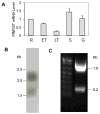
Fig. 1. Transcription of PfMYST gene during parasite intraerythrocytic development
(A) Relative level of PfMYST in parasite by real-time RT-PCR. Constitutively expressed Seryl-tRNA synthetase gene was included as an internal control. Values denote the fold increase in transcription relative to that in the ring stage. R –rings, ET – early trophozoites, LT – late trophozoites, S – schizonts, G – gametocytes. (B) Northern blot analysis of PfMYST expression using total RNA from mixed intraerythrocytic stages. (C) RLM-RACE products showing that the two PfMYST transcripts were initiated at two major positions. (D) Nucleotide sequence at the 5′ and 3′ end of PfMYST. The putative translational start codons (ATGs) of the longer and shorter transcripts are marked as boldface letters. TSSs and polyadenylation sites are indicated by boldface letters and marked with small arrows. Numbering is with respect to the first ATG and stop codon TAA, respectively. Numbers in parentheses indicate the numbers of clones sequenced. A possible polyadenylation signal (AATAAA) is underlined.
Fig. 2
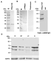
Fig. 2. Expression of PfMYST during parasite development
(A) Purification of 6X His-tagged PfMYST HAT domain (F4) from E. coli. Coomassie-stained SDS-PAGE gel shows the purified PfMYST protein as a 35 kDa band. (B) Western blots of protein extracts from asynchronous 3D7 parasite probed with rabbit preimmune and anti-PfMYST sera. The anti-PfMYST antibodies detected two bands of ~50 and ~70 kDa. (C) Western blot with HRP-conjugated goat anti-rabbit IgG antibodies that bind to the TAP tag. Two bands of 70 and 90 kDa were detected in the TAP-tagged PfMYST clones. (D) Western blot to detect PfMYST protein levels at different stages of the IDC. Equal amounts of proteins (30 μg) from synchronized parasites at ring (R), early trophozoite (ET), late trophozoite (LT) and schizont (S) stages were separated by 12% SDS-PAGE and probed with anti-PfMYST antibodies (upper panel), while protein loading was monitored with the anti-HSP70 antibody (lower panel).
Fig. 3
Fig. 3. Localization of PfMYST
Representative GFP fluorescent images of parasites with GFP fused at the C terminus of the endogenous PfMYST showing the localization of PfMYST-GFP at ring, trophozoite and schizont stages. Nuclei were counterstained with Hoechst 33342. Triple merge indicates the merging of the light, GFP and Hoechst images of the same cells.
Fig. 4
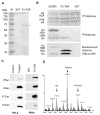
Fig. 4. In vitro acetylation of histones by recombinant PfMYST
(A) Coomassie blue-stained SDS-PAGE gel showing the purification of episomally expressed, TAP-tagged PfMYST F2 fragment (indicated by an asterisk) from transfected parasites (F2-TAP). Wild-type 3D7 parasite serves as a control. (B) In vitro HAT assay showing recombinant PfMYST F2-mediated H4 acetylation of mixed core and nucleosomal histones. Upper panel: Coomassie blue-stained gel; middle panel: fluorography with mixed recombinant P. falciparum core histones as substrates; lower panel: fluorography with histones from HeLa cell mononucleosomes as substrates. (C) Detection of substrate lysine specificity of PfMYST. Left panel represents verification of specificities of the commercial antibodies. Histones isolated from control HeLa cells (Control) and sodium butyrate-treated HeLa cells (Treated) were analyzed by Western blots with antibodies for acetylated lysines H4K5, H4K8, H4K12, and H4K16, respectively. In the right panel, HAT assay was performed with the PfH4 and PfMYST F2-TAP, and acetylated lysines were detected by Western blots with antibodies for acetylated H4K5, K8, K12 and K16. (D) Acetylation of the 24-residue N-terminal H4 peptide by PfMYST F2-TAP was analyzed by mass spectrometry to show mono- (H4-Ac1) and di-acetylated (H4-Ac2) peptides.
Fig. 5
Fig. 5. Deletion analysis to define the active domain of PfMYST
Full-length (F1) PfMYST and seven deletion fragments were episomally expressed in 3D7 parasite as TAP-tagged proteins. The recombinant PfMYST-TAP fragments were purified and used to determine HAT activity in a filter HAT assay. HAT activity was expressed as average amount (CPM) ± standard deviation of 3H incorporated into histones after background correction with wild-type 3D7. The HAT activity of the F1 fragment of PfMYST was set as 100%.
Fig. 6
Fig. 6. Enrichment of PfMYST at the var2csa locus
(A) A simplified scheme of var2csa gene organization showing the TSS and PCR fragment positions (a–h). (B) Distribution of PfMYST along the var2csa gene in CSA selected (var2csaON and var2csaPOISED) and unselected parasites (var2csaOFF) in at ring and mature stages. The relative enrichment of PfMYST at the selected promoter regions (a – h) were determined by ChIP and real-time PCR analysis shown as 2−ΔΔCt value. The results were normalized against ChIP with anti-H4 antibodies for changes in nucleosomal occupancy. Columns labeled with different letters indicate significant difference (P<0.01, Student’s t-test). (C) Ratios of PfMYST enrichment levels along the var2csa gene in CSA-selected versus unselected parasites at ring (filled bars) and mature stages (open bars). Ratios were calculated using relative enrichment levels from 6B. Results are the average of three independent experiments. Error bars denote standard deviations.
Fig. 7
Fig. 7. Overexpression of PfMYST and H4 acetylation
(A) Western blots of PfMYST expression in 3D7-attB parasite with integration of an expression cassette for GFP only (GFP Control), full-length PfMYST-GFP (F1-GFP) and C-terminal truncated PfMYST-GFP (F1C3-GFP) at the cg6 locus. Parasite lysates were probed with anti-PfMYST antibodies and GFP-tagged PfMYST proteins are marked with asterisks. Approximately equal protein loading is indicated with the antibody against HSP70 (lower panel). (B) Effect of PfMYST overexpression on in vivo histone acetylation. Histones were extracted from synchronized parasites at different stages from each parasite line expressing GFP control, F1-GFP and F1C3-GFP. Equal amounts of histone extracts (5 μg/lane) were indicated by Western blot with antibodies against H4. Lysine acetylation was detected by Western blots with antibodies for H4K5ac, H4K8ac, H4K12ac, H4K18ac, and H3K9ac.
Fig. 8
Fig. 8. PfMYST overexpression disturbs parasite cell cycle
(A) Comparison of asexual growth of parasite 3D7-attB, and parasite expressing GFP-control, F1-GFP and F1C3-GFP. Cultures were started at 0.1% parasitemia at ring stage and parasitemia was determined every 48 h for three IDCs. Linear mixed-effects models and Tukey’s pairwise comparison showed that parasitemia between the GFP-control and other PfMYST overexpressing lines were significant at P < 0.05. (B) Comparison of cycle duration of each parasite line: GFP control (46.6 h), F1-GFP (42.8 h), F1C3-GFP (47.3 h). ANOVA analysis of cycle duration showed F1-GFP had significantly shorter cell cycle comparing with other two lines (P < 0.01). (C) Distribution of schizonts with different merozoite numbers for three parasite lines. (D) IDC profile of each parasite line showing the prevalence (percentage) of the ring, trophozoite and schizont stages through a 50 h time period. Note the shortened and prolonged schizont stage for parasite overexpressing F1-GFP and F1C3-GFP, respectively. The star line in F1C3-GFP indicates the presence of schizonts with poorly separated nuclei, which continued to exist in culture at 50–60 h when normal parasites have developed into ring stage.
Fig. 9
Fig. 9. EM comparison of schizonts from control and PfMYST-overexpressing parasites
Representative EM images of 42 h schizonts from GFP-control parasite showing multiple well-separated merozoites, schizonts from F1-GFP parasite with well-separated but fewer merozoites, and abnormal schizonts from F1C3-GFP parasite with less well-separated merozoites (~5% of schizonts). These abnormal schizonts could not complete schizogony and appeared to undergo degradation at later times (at 50 h). Scale bar = 1000 nM.
Fig. 10
Fig. 10. The effect of PfMYST overexpression on parasite DNA replication
DNA content of the parasites was determined using the DNA dye Vybrant Orange and calculated using ring-stage parasite as one genome copy (c). The graphs show the counts of parasites (y axis) with different genome copy numbers (fluorescence intensities) (x-axis). Parasites were collected at trophozoite (30 h), late trophozoite (36 h) and schizont (42 h) stages.

Similar articles

Cited by

References

    1. Adachi N, Kimura A, Horikoshi M. A conserved motif common to the histone acetyltransferase Esa1 and the histone deacetylase Rpd3. J Biol Chem. 2002;277:35688–35695. - PubMed
    1. Akhtar A, Becker PB. The histone H4 acetyltransferase MOF uses a C2HC zinc finger for substrate recognition. EMBO Reports. 2001;2:113–118. - PMC - PubMed
    1. Akhtar A, Zink D, Becker PB. Chromodomains are protein-RNA interaction modules. Nature. 2000;407:405–409. - PubMed
    1. Alkhalil A, Achur RN, Valiyaveettil M, Ockenhouse CF, Gowda DC. Structural requirements for the adherence of Plasmodium falciparum-infected erythrocytes to chondroitin sulfate proteoglycans of human placenta. J Biol Chem. 2000;275:40357–40364. - PubMed
    1. Andrews KT, Tran TN, Lucke AJ, Kahnberg P, Le GT, Boyle GM, et al. Potent antimalarial activity of histone deacetylase inhibitor analogues. Antimicrol Agents Chemother. 2008;52:1454–1461. - PMC - PubMed

Publication types

MeSH terms