Congenic strains provide evidence that a mapped locus on chromosome 15 influences excitotoxic cell death
- PMID: 20807240
- PMCID: PMC3005149
- DOI: 10.1111/j.1601-183X.2010.00644.x
Congenic strains provide evidence that a mapped locus on chromosome 15 influences excitotoxic cell death
Abstract
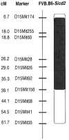
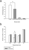
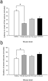
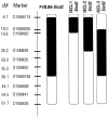
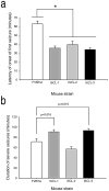
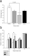
Inbred strains of mice differ in their susceptibility to excitotoxin-induced cell death, but the genetic basis of individual variation is unknown. Prior studies with crosses of the FVB/NJ (seizure-induced cell death susceptible) mouse and the seizure-induced cell death resistant mouse, C57BL/6J, showed the presence of three quantitative trait loci (QTLs), named seizure-induced cell death 1 (Sicd1) to Sicd3. To better localize and characterize the Sicd2 locus, two reciprocal congenic mouse strains were created. While the B6.FVB-Sicd2 congenic mouse was without effect on modifying susceptibility to seizure-induced excitotoxic cell death, the FVB.B6-Sicd2 congenic mouse, in which the chromosome (Chr) 15 region of C57BL/6J was introgressed into FVB/NJ, showed reduced seizure-induced excitotoxic cell death following kainate administration. Phenotypic comparison between FVB and the congenic FVB.B6-Sicd2 strain confirmed that the Sicd2 interval harbors gene(s) conferring strong protection against seizure-induced excitotoxic cell death. Interval-specific congenic lines (ISCLs) that encompass Sicd2 on Chr 15 were generated and were used to fine-map this QTL. Resultant progeny were treated with kainate and examined for the extent of seizure-induced cell death in order to deduce the Sicd2 genotypes of the recombinants through linkage analysis. All of the ISCLs exhibited reduced cell death associated with the C57BL/6J phenotype; however, ISCL-2 showed the most dramatic reduction in seizure-induced cell death in both area CA3 and in the dentate hilus. These findings confirm the existence of polymorphic loci within the reduced critical region of Sicd2 that regulate the severity of seizure-induced cell death.
© 2010 The Author. Genes, Brain and Behavior © 2010 Blackwell Publishing Ltd and International Behavioural and Neural Genetics Society.
Figures


Similar articles
-
Susceptibility to seizure-induced excitotoxic cell death is regulated by an epistatic interaction between Chr 18 (Sicd1) and Chr 15 (Sicd2) loci in mice.PLoS One. 2014 Oct 15;9(10):e110515. doi: 10.1371/journal.pone.0110515. eCollection 2014. PLoS One. 2014. PMID: 25333963 Free PMC article.
-
Microarray-assisted fine mapping of quantitative trait loci on chromosome 15 for susceptibility to seizure-induced cell death in mice.Eur J Neurosci. 2013 Dec;38(12):3679-90. doi: 10.1111/ejn.12351. Epub 2013 Sep 3. Eur J Neurosci. 2013. PMID: 24001120 Free PMC article.
-
A quantitative trait locus on chromosome 18 is a critical determinant of excitotoxic cell death susceptibility.Eur J Neurosci. 2007 Apr;25(7):1998-2008. doi: 10.1111/j.1460-9568.2007.05443.x. Eur J Neurosci. 2007. PMID: 17439488
-
Variation in Galr1 expression determines susceptibility to exocitotoxin-induced cell death in mice.Genes Brain Behav. 2008 Jul;7(5):587-98. doi: 10.1111/j.1601-183X.2008.00395.x. Genes Brain Behav. 2008. PMID: 18363852 Free PMC article.
-
A novel strategy for genetic dissection of complex traits: the population of specific chromosome substitution strains from laboratory and wild mice.Mamm Genome. 2010 Aug;21(7-8):370-6. doi: 10.1007/s00335-010-9270-x. Epub 2010 Jul 11. Mamm Genome. 2010. PMID: 20623355 Review.
Cited by
-
Susceptibility to seizure-induced excitotoxic cell death is regulated by an epistatic interaction between Chr 18 (Sicd1) and Chr 15 (Sicd2) loci in mice.PLoS One. 2014 Oct 15;9(10):e110515. doi: 10.1371/journal.pone.0110515. eCollection 2014. PLoS One. 2014. PMID: 25333963 Free PMC article.
-
The relevance of individual genetic background and its role in animal models of epilepsy.Epilepsy Res. 2011 Nov;97(1-2):1-11. doi: 10.1016/j.eplepsyres.2011.09.005. Epub 2011 Oct 15. Epilepsy Res. 2011. PMID: 22001434 Free PMC article. Review.
-
A locus on mouse Ch10 influences susceptibility to limbic seizure severity: fine mapping and in silico candidate gene analysis.Genes Brain Behav. 2014 Mar;13(3):341-9. doi: 10.1111/gbb.12116. Epub 2014 Jan 27. Genes Brain Behav. 2014. PMID: 24373497 Free PMC article.
-
Lack of Chronic Histologic Lesions Supportive of Sublethal Spontaneous Seizures in FVB/N Mice.Comp Med. 2016 Apr;66(2):105-11. Comp Med. 2016. PMID: 27053564 Free PMC article.
-
Microarray-assisted fine mapping of quantitative trait loci on chromosome 15 for susceptibility to seizure-induced cell death in mice.Eur J Neurosci. 2013 Dec;38(12):3679-90. doi: 10.1111/ejn.12351. Epub 2013 Sep 3. Eur J Neurosci. 2013. PMID: 24001120 Free PMC article.
References
-
- Ben-Ari Y. Limbic seizure and brain damage produced by kainic acid: mechanisms and relevance to human temporal lobe epilepsy. Neuroscience. 1985;14:375–403. - PubMed
-
- Ben-Ari Y, Cossart R. Kainate, a double agent that generates seizures: two decades of progress. Trends Neurosci. 2000;23:580–587. - PubMed
-
- Bennett B, Carosone-Link P, Beeson M, Gordon L, Phares-Zook N, Johnson TE. Genetic dissection of quantitative trait locus for ethanol sensitivity in long- and short-sleep mice. Genes Brain Behav. 2008;7:659–668. - PubMed
-
- Berger ML, Lefauconnier JM, Tremblay E, Ben-Ari Y. Limbic seizures induced by systemically applied kainic acid: how much kainic acid reaches the brain? Adv Exp Med Biol. 1986;203:199–209. - PubMed
MeSH terms
Substances
Grants and funding
LinkOut - more resources
Full Text Sources
Molecular Biology Databases
Miscellaneous

