. 2010 Nov 1;347(1):204-15.
doi: 10.1016/j.ydbio.2010.08.025. Epub 2010 Aug 31.

Crossveinless-2 is required for the relocalization of Chordin protein within the vertebral field in mouse embryos

Affiliations

Crossveinless-2 is required for the relocalization of Chordin protein within the vertebral field in mouse embryos

Lise Zakin et al. Dev Biol. .

Abstract

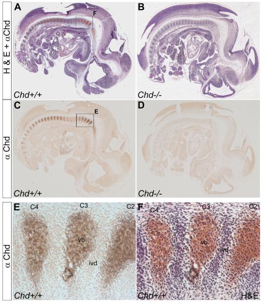
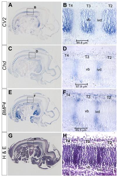
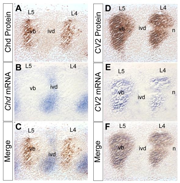
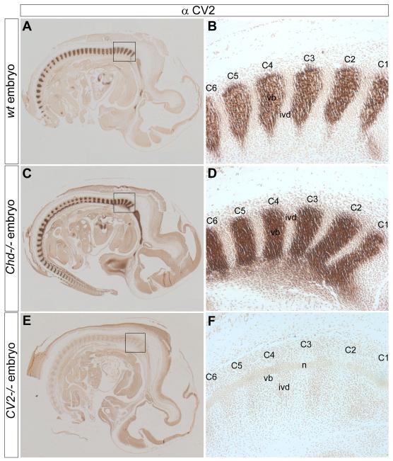
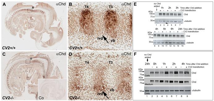
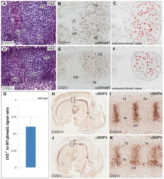
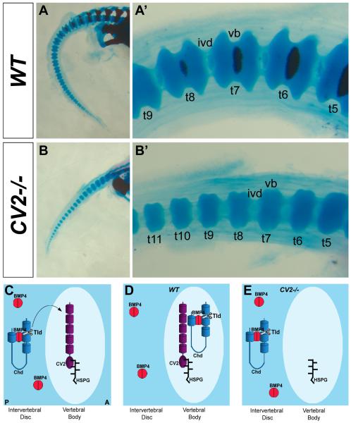
Bone morphogenetic proteins (BMPs), as well as the BMP-binding molecules Chordin (Chd), Crossveinless-2 (CV2) and Twisted Gastrulation (Tsg), are essential for axial skeletal development in the mouse embryo. We previously reported a strong genetic interaction between CV2 and Tsg and proposed a role for this interaction in the shaping of the BMP morphogenetic field during vertebral development. In the present study we investigated the roles of CV2 and Chd in the formation of the vertebral morphogenetic field. We performed immunostainings for CV2 and Chd protein on wild-type, CV2(-/-) or Chd(-/-) mouse embryo sections at the stage of onset of the vertebral phenotypes. By comparing mRNA and protein localizations we found that CV2 does not diffuse away from its place of synthesis, the vertebral body. The most interesting finding of this study was that Chd synthesized in the intervertebral disc accumulates in the vertebral body. This relocalization does not take place in CV2(-/-) mutants. Instead, Chd was found to accumulate at its site of synthesis in CV2(-/-) embryos. These results indicate a CV2-dependent flow of Chd protein from the intervertebral disc to the vertebral body. Smad1/5/8 phosphorylation was decreased in CV2(-/-)vertebral bodies. This impaired BMP signaling may result from the decreased levels of Chd/BMP complexes diffusing from the intervertebral region. The data indicate a role for CV2 and Chd in the establishment of the vertebral morphogenetic field through the long-range relocalization of Chd/BMP complexes. The results may have general implications for the formation of embryonic organ-forming morphogenetic fields.


Figures

Fig. 1
Fig. 1
Chd protein concentrates in the prospective vertebral bodies. (A) Sagittal section of a Wild-type 12.5 d.p.c. mouse embryo stained with Chd antibody and then counterstained with H&E. (B) Sagittal section of a 12.5 d.p.c. Chd−/− mouse embryo similarly stained. (C) Chd antibody staining on wild-type embryo, showing Chd protein localized in prospective vertebral body. (D) No signal is detected on Chd−/− mouse embryo section, using Chd antibody thus showing that the Chd antibody is specific (n=2 Chd−/− embryos, with similar results). (E) Magnified view of boxed area in (C) showing Chd staining in vertebral bodies C2 through C4. (F) Same section as in (E) counterstained with H&E. C2-C4, cervical vertebrae; ivd, prospective intervertebral disc; vb, prospective vertebral body.
Fig. 2
Fig. 2
CV2 mRNA is transcribed in the prospective vertebral bodies, while Chd and BMP4 mRNAs are expressed in the prospective intervertebral discs. In situ hybridizations and H&E staining were performed on adjacent sagittal sections of the same 12.5 d.p.c. wild-type mouse embryo. (A) CV2 in situ hybridization, showing CV2 mRNA localized in the prospective vertebral bodies. (B) Magnified view of area boxed in (A) showing CV2 staining in thoracic vertebrae (T) 2 through 4. (C) Chd in situ hybridization showing Chd mRNA expressed in prospective intervertebral disc. (D) Magnified view of area boxed in (C) showing Chd mRNA expression. (E) BMP4 in situ hybridization showing BMP4 mRNA expression in prospective intervertebral discs. (F) Magnified view of area boxed in (E) showing BMP4 mRNA staining. (G) H&E staining on a section adjacent to the one shown in (E). (H) Magnified view of area boxed in (G) showing regions of low and high tissue density corresponding to the prospective vertebral body and intervertebral disc, respectively. The bars in B, D and F indicate the distance in μm between the intervertebral disc and the center of the vertebral body.
Fig. 3
Fig. 3
Chd mRNA is synthesized in the prospective intervertebral disc while Chd protein accumulates in the prospective vertebral bodies, in which CV2 mRNA and protein are also found. In situ hybridizations and immunostainings were performed on adjacent sagittal sections of a 12.5 d.p.c. wild-type mouse embryo. (A) Chd antibody staining showing Chd protein localized to the prospective lumbar (L) vertebral bodies. (B) Chd in situ hybridization in adjoining section showing Chd mRNA expressed in prospective intervertebral discs. (C) Merged image of panels (A) and (B), showing that Chd mRNA and Chd protein are localized in complementary regions. Chd protein accumulates at a considerable distance from where it is synthesized. Note also that Chd accumulates predominantly in the posterior half of the vertebral body at this lumbar level. (D) CV2 antibody staining showing CV2 protein localized in the prospective vertebral body. (E) CV2 in situ hybridization showing CV2 mRNA localized in the prospective vertebral body. (F) Merged images showing that CV2 mRNA and CV2 protein co-localize. The secreted CV2 protein does not diffuse far from its site of synthesis. n, notochord.
Fig. 4
Fig. 4
The localization of CV2 is independent of Chd. Immunostainings with a CV2 antibody performed on sagittal sections of 12.5 d.p.c. wild-type, Chd−/− and CV2−/− mouse embryos. (A) CV2 antibody staining on a wild-type section, showing the presence of CV2 in the prospective vertebral body. (B) Magnified view of area boxed in (A) showing staining in the prospective vertebral bodies of cervical vertebrae C1 through C6. (C) CV2 antibody staining is normal in Chd−/− embryos. The basioccipital bone is also stained anterior to the vertebral column. (D) Higher-power view of area boxed area in (C) showing staining of CV2 in C1 through C6 similar to that of the wild-type embryo in panel (B). In this Chd−/− animal, CV2 staining was stronger than wild-type (compare panels A and C); however, this difference was not observed in two other mutants analyzed. (E) CV2 antibody staining was undetectable on a CV2−/− section, demonstrating the specificity of the antibody. (F) Magnified view of boxed area in (E).
Fig. 5
Fig. 5
In the mouse embryo, Chd protein requires CV2 for its accumulation in the prospective vertebral bodies, and in cultured cells CV2 serves as a sink for Chd. Immunostainings with a Chd antibody and H&E staining performed on sagittal sections of 12.5 d.p.c. wild-type and CV2−/− embryos. (A) Chd antibody staining on a wild-type embryo, showing the accumulation of Chd in prospective vertebral bodies (vb). (B) Magnified view of area boxed in (A). (C) Chd antibody staining on CV2−/− embryo, showing that Chd protein accumulation is not detectable in vertebral bodies in the absence of CV2. (D) Magnified views of area boxed in (C). Note that mutation of CV2 prevents Chd accumulation in the prospective vertebral bodies; this result was observed in 4 different CV2 mutant embryos (8 independent experiments). (E) Western blot using an anti-Chd antibody showing that overexpression of a full-length CV2 construct in Cos7 cells does not stabilize added recombinant Chd over the course of 24 hours. Samples containing culture medium and cell extracts were collected at different time points; tubulin was used as a loading control. Note that CV2 does not affect Chd stability. (F) Western blot analysis of the cell fraction (containing cells and extracellular matrix) or culture medium from Cos7 cells minus or plus CV2 overexpression and treated with recombinant mouse Chd. Note that already after 1 hour of addition of recombinant Chd protein, CV2 is able to sequester Chd.
Fig. 6
Fig. 6
Localization and quantification of Smad1/5/8 phosphorylation and BMP4 expression in the vertebral column of wild-type and CV2−/− embryos. H&E staining, α–pSmad1/5/8 and α–BMP4 immunostainings were performed on sagittal sections of 12.5 d.p.c. mouse embryos. (A, D) H&E staining showing a similar cell density in the vertebral bodies (vb) of wild-type and CV2−/− embryos, even though the size of the vertebral bodies appears reduced and the intervertebral disc (ivd) domain is expanded in the mutant. (B, E) pSmad1/5/8 signal is nuclear in the vertebral bodies of wild-type and CV2−/− embryos. (C, F) Quantification of the nuclear pSmad1/5/8 signal. Pixels above background level in the outlined vertebral bodies are shown in red. Quantifications of 6 vertebral bodies indicated that CV2−/− cartilage precursors were less stained than in wild-type. (G) Quantification showing that the average ratio of pSmad1/5/8 signal between CV2−/− and wild-type was 60%. The error bar corresponds to the standard deviation of the ratio of pSmad1/5/8 signal between different individual vertebral bodies. The wild-type value is 1 as indicated. (H, J) BMP4 antibody staining on wild-type and CV2−/− embryos showing the presence of BMP4 protein in prospective intervertebral discs (ivd). (I, K) Higher power view of thoracic vertebrae 4 (T4) and 5 (T5) from sections shown in (H) and (J), respectively, showing a broadening of the BMP4 expression domain in the mutant reflecting the widening of the intervertebral disc region.
Fig. 7
Fig. 7
The CV2−/− phenotype in vertebral body cartilage and model of the role of CV2 in the proper localization of Chd in the mouse vertebral field. (A-B’) Skeletal preparations of wild-type (top panels) and CV2−/− (bottom panels) neonates stained with Alcian Blue (cartilage) and Alizarin Red (bone). Tail area is shown at low (left panels) and high (right panels) power magnifications. Note in the vertebral body cartilage of CV2−/− the reduction in size and loss of the spool-like shape compared to the wild-type. We propose that this phenotype is explained in part by the lack of facilitated diffusion of Chd/Tsg/BMP4 complexes from the intervertebral disc to the vertebral body. (C) Model depicting the localization of Chd, CV2 and BMP4 in wild-type and CV2−/− developing vertebrae. In the wild-type CV2 is retained in the vertebral body through its interaction with heparin sulfate proteoglycans (HSPGs), which are very abundant in developing cartilage matrix. (D) Chd/BMP4 complexes diffuse from the intervertebral disc, where they are synthesized, to the vertebral body where they bind to CV2. Once in the vertebral body, Chd is cleaved by a member of the Tolloid metalloproteinase family (Tld) allowing the release of BMP4 in the vertebral bodies so that the high BMP signaling levels required for cartilage development are reached. (E) In CV2−/− vertebrae, Chd/BMP4 complexes do not flow from the intervertebral disc to the vertebral body but instead remain in the intervertebral disc, where they are degraded by the action of Tolloid metalloproteinases on Chd. A, anterior; P, posterior; t5-t11 numbered tail vertebrae.

Similar articles

Cited by

References

    1. Ambrosio AL, Taelman VF, Lee HX, Metzinger CA, Coffinier C, De Robertis EM. Crossveinless-2 Is a BMP feedback inhibitor that binds Chordin/BMP to regulate Xenopus embryonic patterning. Dev. Cell. 2008;15:248–260. - PMC - PubMed
    1. Bachiller D, Klingensmith J, Kemp C, Belo JA, Anderson RM, May SR, McMahon JA, McMahon AP, Harland RM, Rossant J, De Robertis EM. The organizer factors Chordin and Noggin are required for mouse forebrain development. Nature. 2000;403:658–661. - PubMed
    1. Bachiller D, Klingensmith J, Shneyder N, Tran U, Anderson R, Rossant J, De Robertis EM. The role of Chordin/BMP signals in mammalian pharyngeal development and DiGeorge syndrome. Development. 2003;130:3567–3578. - PubMed
    1. Bagnall KM, Higgins SJ, Sanders EJ. The contribution made by a single somite to the vertebral column: experimental evidence in support of resegmentation using the chick-quail chimaera model. Development. 1988;103:69–85. - PubMed
    1. Ben-Zvi D, Shilo BZ, Fainsod A, Barkai N. Scaling of the BMP activation gradient in Xenopus embryos. Nature. 2008;453:1205–1211. - PubMed

Publication types

MeSH terms