Changes in and dorsal to the rat suprachiasmatic nucleus during early pregnancy
- PMID: 20807562
- PMCID: PMC2975742
- DOI: 10.1016/j.neuroscience.2010.08.057
Changes in and dorsal to the rat suprachiasmatic nucleus during early pregnancy
Abstract
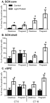
Circadian rhythms in behavior and physiology change as female mammals transition from one reproductive state to another. The mechanisms responsible for this plasticity are poorly understood. The suprachiasmatic nucleus (SCN) of the hypothalamus contains the primary circadian pacemaker in mammals, and a large portion of its efferent projections terminate in the ventral subparaventricular zone (vSPZ), which also plays important roles in rhythm regulation. To determine whether these regions might mediate changes in overt rhythms during early pregnancy, we first compared rhythms in Fos and Per2 protein expression in the SCN and vSPZ of diestrous and early pregnant rats maintained in a 12:12-h light/dark (LD) cycle. No differences in the Fos rhythm were seen in the SCN core, but in the SCN shell, elevated Fos expression was maintained throughout the light phase in pregnant, but not diestrous, rats. In the vSPZ, the Fos rhythm was bimodal in diestrous rats, but this rhythm was lost in pregnant rats. Peak Per2 expression was phase-advanced by 4 h in the SCN of pregnant rats, and some differences in Per2 expression were found in the vSPZ as well. To determine whether differences in Fos expression were due to altered responsivity to light, we next characterized light-induced Fos expression in the SCN and vSPZ of pregnant and diestrous rats in the mid-subjective day and night. We found that the SCN core of the two groups responded in the same way at each time of day, whereas the rhythm of Fos responsivity in the SCN shell and vSPZ differed between diestrous and pregnant rats. These results indicate that the SCN and vSPZ are functionally re-organized during early pregnancy, particularly in how they respond to the photic environment. These changes may contribute to changes in overt behavioral and physiological rhythms that occur at this time.
Copyright © 2010 IBRO. Published by Elsevier Ltd. All rights reserved.
Figures


Similar articles
-
Site-specific changes in brain extra-SCN oscillators during early pregnancy in the rat.J Biol Rhythms. 2011 Aug;26(4):363-7. doi: 10.1177/0748730411409714. J Biol Rhythms. 2011. PMID: 21775295
-
Expression profiles of PER2 immunoreactivity within the shell and core regions of the rat suprachiasmatic nucleus: lack of effect of photic entrainment and disruption by constant light.J Mol Neurosci. 2003;21(2):133-47. doi: 10.1385/JMN:21:2:133. J Mol Neurosci. 2003. PMID: 14593213
-
Differences in the suprachiasmatic nucleus and lower subparaventricular zone of diurnal and nocturnal rodents.Neuroscience. 2004;127(1):13-23. doi: 10.1016/j.neuroscience.2004.04.049. Neuroscience. 2004. PMID: 15219664
-
Temporal and spatial distribution of immunoreactive PER1 and PER2 proteins in the suprachiasmatic nucleus and peri-suprachiasmatic region of the diurnal grass rat (Arvicanthis niloticus).Brain Res. 2006 Feb 16;1073-1074:348-58. doi: 10.1016/j.brainres.2005.11.082. Epub 2006 Jan 20. Brain Res. 2006. PMID: 16430875
-
Individual differences and diversity in human physiological responses to light.EBioMedicine. 2022 Jan;75:103640. doi: 10.1016/j.ebiom.2021.103640. Epub 2022 Jan 10. EBioMedicine. 2022. PMID: 35027334 Free PMC article. Review.
Cited by
-
Impact of chronodisruption during primate pregnancy on the maternal and newborn temperature rhythms.PLoS One. 2013;8(2):e57710. doi: 10.1371/journal.pone.0057710. Epub 2013 Feb 28. PLoS One. 2013. PMID: 23469055 Free PMC article.
-
Light stimulates the mouse adrenal through a retinohypothalamic pathway independent of an effect on the clock in the suprachiasmatic nucleus.PLoS One. 2014 Mar 21;9(3):e92959. doi: 10.1371/journal.pone.0092959. eCollection 2014. PLoS One. 2014. PMID: 24658072 Free PMC article.
-
Does Circadian Disruption Play a Role in the Metabolic-Hormonal Link to Delayed Lactogenesis II?Front Nutr. 2015 Feb 23;2:4. doi: 10.3389/fnut.2015.00004. eCollection 2015. Front Nutr. 2015. PMID: 25988133 Free PMC article. Review.
-
Antibodies for assessing circadian clock proteins in the rodent suprachiasmatic nucleus.PLoS One. 2012;7(4):e35938. doi: 10.1371/journal.pone.0035938. Epub 2012 Apr 27. PLoS One. 2012. PMID: 22558277 Free PMC article.
-
Circadian clocks and their integration with metabolic and reproductive systems: our current understanding and its application to the management of dairy cows.J Anim Sci. 2022 Oct 1;100(10):skac233. doi: 10.1093/jas/skac233. J Anim Sci. 2022. PMID: 35775472 Free PMC article. Review.
References
-
- Abizaid A, Mezei G, Horvath TL. Estradiol enhances light-induced expression of transcription factors in the SCN. Brain Res. 2004;1010:35–44. - PubMed
-
- Abrahamson EE, Moore RY. Lesions of suprachiasmatic nucleus efferents selectively affect rest-activity rhythm. Mol Cell Endocrinol. 2006;252:46–56. - PubMed
-
- Albers HE. Gonadal hormones organize and modulate the circadian system of the rat. Am J Physiol Regul Integr Comp Physiol. 1981;241:62–66. - PubMed
-
- Albers HE, Gerall AA, Axelson JF. Effect of reproductive state on circadian periodicity in the rat. Physiol Behav. 1981;26:21–25. - PubMed
-
- Antle MC, Silver R. Orchestrating time: arrangements of the brain circadian clock. Trends Neurosci. 2005;28:145–151. - PubMed
Publication types
MeSH terms
Substances
Grants and funding
LinkOut - more resources
Full Text Sources
Research Materials

