. 2011 Jan;23(1):135-44.
doi: 10.1016/j.cellsig.2010.08.013. Epub 2010 Aug 31.

Activation of the JNK signalling pathway by macrophage migration inhibitory factor (MIF) and dependence on CXCR4 and CD74

Affiliations

Activation of the JNK signalling pathway by macrophage migration inhibitory factor (MIF) and dependence on CXCR4 and CD74

Hongqi Lue et al. Cell Signal. 2011 Jan.

Abstract

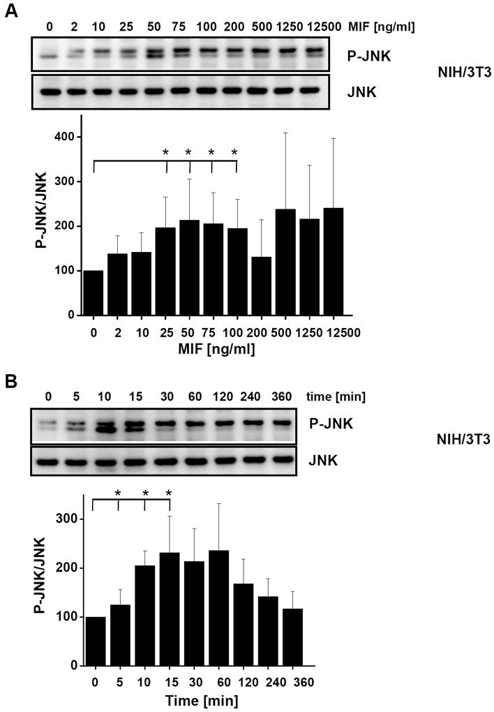
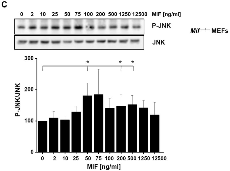
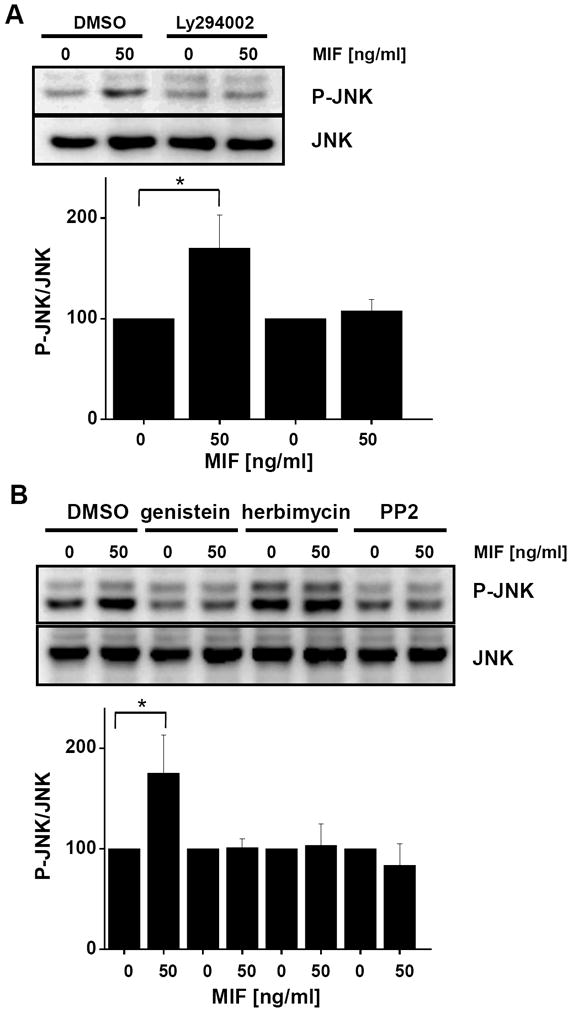
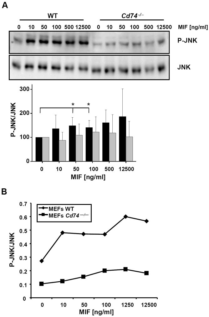
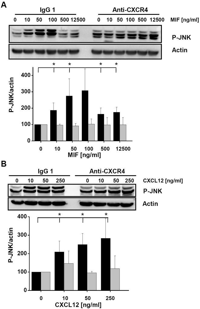
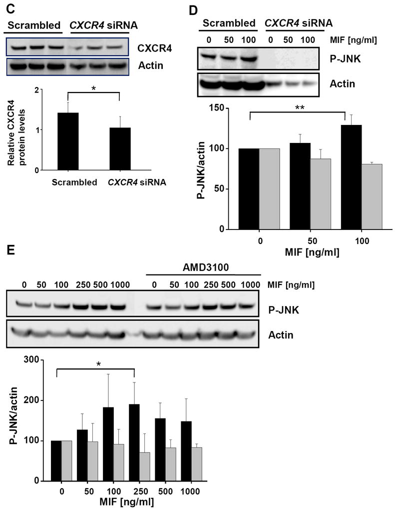
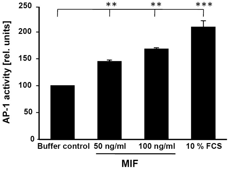
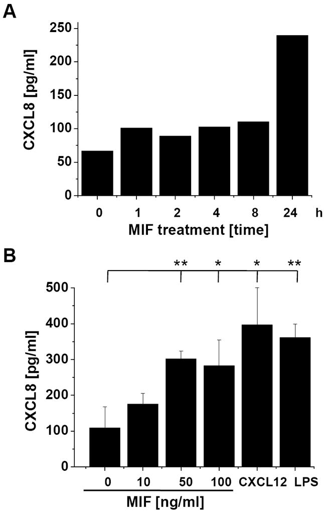
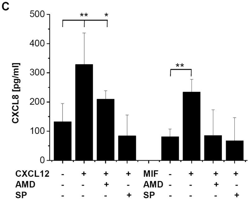
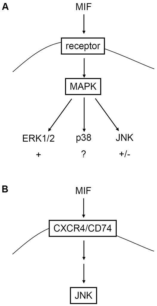
c-Jun N-terminal kinase (JNK) is a member of the mitogen-activated protein kinase (MAPK) family and controls essential processes such as inflammation, cell differentiation, and apoptosis. JNK signalling is triggered by extracellular signals such as cytokines and environmental stresses. Macrophage migration inhibitory factor (MIF) is a pleiotropic pro-inflammatory cytokine with chemokine-like functions in leukocyte recruitment and atherosclerosis. MIF promotes MAPK signalling through ERK1/2, while it can either activate or inhibit JNK phosphorylation, depending on the cell type and underlying stimulation context. MIF activities are mediated by non-cognate interactions with the CXC chemokine receptors CXCR2 and CXCR4 or by ligation of CD74, which is the cell surface expressed form of the class II invariant chain. ERK1/2 signalling stimulated by MIF is dependent on CD74, but the receptor pathway involved in MIF activation of the JNK pathway is unknown. Here we comprehensively characterize the stimulatory effect of MIF on the canonical JNK/c-Jun/AP-1 pathway in fibroblasts and T cell lines and identify the upstream signalling components. Physiological concentrations of recombinant MIF triggered the phosphorylation of JNK and c-Jun and rapidly activated AP-1. In T cells, MIF-mediated activation of the JNK pathway led to upregulated gene expression of the inflammatory chemokine CXCL8. Activation of JNK signalling by MIF involved the upstream kinases PI3K and SRC and was found to be dependent on CXCR4 and CD74. Together, these data show that the CXCR4/CD74/SRC/PI3K axis mediates a rapid and transient activation of the JNK pathway as triggered by the inflammatory cytokine MIF in T cells and fibroblasts.


Figures

Fig. 1
Fig. 1
MIF promotes rapid phosphorylation of c-Jun. MIF promotes rapid and transient phosphorylation of c-Jun in NIH/3T3 fibroblasts. A time course from 0-180 min is shown. Phospho-c-Jun levels were determined by Western blot analysis and c-Jun expression levels were used for standardization. The blot shown is representative of three independent experiments.
Fig. 2
Fig. 2
MIF promotes JNK activation in fibroblasts in a concentration- and time-dependent manner. (A) Concentration-dependent phosphorylation of JNK by rMIF in NIH/3T3 fibroblasts. Maximum activation occurs at 50 ng/ml MIF. Upper panel, JNK phosphorylation was measured by Western blot analysis using a pan-phospho-JNK antibody. Lower panel, band densitometry was performed from 4 independent experiments (means ± SD) to quantify MIF-mediated JNK phosphorylation using JNK levels for normalization. (B) JNK phosphorylation by MIF is time-dependent and transient and peaks at an early time point of 5-15 min. MIF-mediated JNK phosphorylation was measured and quantified as in (A). Data are means ± SD from three independent experiments. (C) Concentration-dependent phosphorylation of JNK by rMIF in mouse embryonal fibroblasts genetically deficient for MIF (Mif −/− MEFs). Maximum activation starts at 50 ng/ml MIF. Quantification of JNK activation as in (A). Data are means ± SD from three independent experiments. Asterisks (*) indicate statistical significance (p<0.05) between the control value (0 ng/ml MIF or 0 min) and the corresponding incubations.
Fig. 2
Fig. 2
MIF promotes JNK activation in fibroblasts in a concentration- and time-dependent manner. (A) Concentration-dependent phosphorylation of JNK by rMIF in NIH/3T3 fibroblasts. Maximum activation occurs at 50 ng/ml MIF. Upper panel, JNK phosphorylation was measured by Western blot analysis using a pan-phospho-JNK antibody. Lower panel, band densitometry was performed from 4 independent experiments (means ± SD) to quantify MIF-mediated JNK phosphorylation using JNK levels for normalization. (B) JNK phosphorylation by MIF is time-dependent and transient and peaks at an early time point of 5-15 min. MIF-mediated JNK phosphorylation was measured and quantified as in (A). Data are means ± SD from three independent experiments. (C) Concentration-dependent phosphorylation of JNK by rMIF in mouse embryonal fibroblasts genetically deficient for MIF (Mif −/− MEFs). Maximum activation starts at 50 ng/ml MIF. Quantification of JNK activation as in (A). Data are means ± SD from three independent experiments. Asterisks (*) indicate statistical significance (p<0.05) between the control value (0 ng/ml MIF or 0 min) and the corresponding incubations.
Fig. 3
Fig. 3
MIF-stimulated JNK phosphorylation is dependent on the upstream activation of PI3K and SRC-type kinases. (A) Activation of transient JNK phosphorylation by MIF is inhibited by the PI3K inhibitor Ly294002. NIH/3T3 fibroblasts were incubated with rMIF for 10 min in the presence of Ly294002 or solvent control (DMSO). Upper panel: JNK phosphorylation was measured by Western blot analysis using a pan-phospho-JNK antibody; lower panel: Statistical analysis of the blots was performed by band densitometry to quantify MIF-mediated JNK phosphorylation using JNK levels for normalization (means ± SD of three independent experiments). (B) MIF-induced JNK phosphorylation in fibroblasts is inhibited by the broad spectrum tyrosine kinase inhibitor genistein or the SRC kinase-specific inhibitors herbimycin or PP2. NIH/3T3 cells were stimulated with rMIF for 10 min in the presence of genistein, herbimycin, PP2, or solvent control (DMSO). Western blot (upper panel) and statistical analysis (lower panel; means ± SD of three independent experiments) was performed as in (A).
Fig. 4
Fig. 4
MIF-induced JNK phosphorylation is partially dependent on CD74. (A) Phospho-JNK Western blot of lysates from MIF-stimulated wildtype (WT) and Cd74−/− MEFs. Various concentrations of rMIF were added as indicated. JNK phosphorylation was measured by phospho-JNK-specific Western blot (upper panel) and quantified as before (lower panel; means ± SD of 5 independent experiments). (B) Line graph correlating the phosphorylation ratios as obtained in (A) over the concentration of rMIF and comparison between the effect of MIF on JNK phosphorylation in WT versus Cd74−/− MEFs.
Fig. 5
Fig. 5
JNK activation stimulated by MIF is dependent on CXCR4. (A) Activation of JNK phosphorylation by MIF in Jurkat T cells is abolished by anti-CXCR4 antibodies. Jurkat cells were incubated with various concentrations of rMIF for 10 min in the presence of anti-CXCR4 or isotype control antibodies. A quantification of MIF-induced JNK phosphorylation was determined by Western blotting using a pan-phospho-JNK antibody and actin for normalization (upper panel) and statistical analysis of 4 independent experiments (means ± SD; lower panel) as before. (B) Activation of JNK phosphorylation by CXCL12 in Jurkat T cells is blocked by anti-CXCR4 antibodies; the data shown are means ± SD from 4 independent experiments; otherwise, see under (A). (C) Knock down of CXCR4 in Jurkat T cells by siRNA-based gene silencing. Top panel, Western blot against CXCR4 from lysates of Jurkat cells treated with CXCR4 siRNA or scrambled control RNA. Actin was used for standardization. Knock down data are from 4 independent experiments. Bottom panel, quantification by band densitometry using actin as standard (bars represent means ± SD of n=4 experiments). (D) Knock down of CXCR4 abolishes phosphorylation of JNK by MIF in Jurkat cells. Comparison between the effect of CXCR4 siRNA and scrambled RNA (upper panel: Western blot using a pan-phospho-JNK antibody and actin as a standard; lower panel: statistical analysis of 4 independent experiments; means ± SD). (E) The CXCR4-specific inhibitor AMD3100 blocks MIF-induced JNK phosphorylation. Jurkat T cells were treated with indicated concentrations of rMIF in the presence or absence of 1 μg/ml AMD3100. Analysis as before (means ± SD of three independent experiments each). Asterisks indicate statistical significance: * p<0.05; ** p<0.01.
Fig. 5
Fig. 5
JNK activation stimulated by MIF is dependent on CXCR4. (A) Activation of JNK phosphorylation by MIF in Jurkat T cells is abolished by anti-CXCR4 antibodies. Jurkat cells were incubated with various concentrations of rMIF for 10 min in the presence of anti-CXCR4 or isotype control antibodies. A quantification of MIF-induced JNK phosphorylation was determined by Western blotting using a pan-phospho-JNK antibody and actin for normalization (upper panel) and statistical analysis of 4 independent experiments (means ± SD; lower panel) as before. (B) Activation of JNK phosphorylation by CXCL12 in Jurkat T cells is blocked by anti-CXCR4 antibodies; the data shown are means ± SD from 4 independent experiments; otherwise, see under (A). (C) Knock down of CXCR4 in Jurkat T cells by siRNA-based gene silencing. Top panel, Western blot against CXCR4 from lysates of Jurkat cells treated with CXCR4 siRNA or scrambled control RNA. Actin was used for standardization. Knock down data are from 4 independent experiments. Bottom panel, quantification by band densitometry using actin as standard (bars represent means ± SD of n=4 experiments). (D) Knock down of CXCR4 abolishes phosphorylation of JNK by MIF in Jurkat cells. Comparison between the effect of CXCR4 siRNA and scrambled RNA (upper panel: Western blot using a pan-phospho-JNK antibody and actin as a standard; lower panel: statistical analysis of 4 independent experiments; means ± SD). (E) The CXCR4-specific inhibitor AMD3100 blocks MIF-induced JNK phosphorylation. Jurkat T cells were treated with indicated concentrations of rMIF in the presence or absence of 1 μg/ml AMD3100. Analysis as before (means ± SD of three independent experiments each). Asterisks indicate statistical significance: * p<0.05; ** p<0.01.
Fig. 6
Fig. 6
Rapid activation of the AP-1 transcription factor by MIF. HEK293 cells were treated with indicated concentrations of rMIF for 10 min, nuclear extracts isolated and analyzed by AP-1/promoter-specific ELISA using the Trans-AM AP-1 kit with an immobilized consensus oligonucleotide containing the TPA response element (TRE) site (5′-TGAGTCA-3′). Relative AP-1 activity units are shown determined by ELISA using a phospho-c-Jun specific antibody (Ser-73) and HRP-conjugated secondary antibody and colorimetric development (450 nm). Values shown were normalized over the protein concentration of the nuclear extracts and represent means ± SD of triplicate measurements. Asterisks indicate statistical significance: ** p<0.01; *** p<0.005.
Fig. 7
Fig. 7
MIF-induced CXCL8 secretion from Jurkat T cells is dependent on CXCR4 and JNK. (A) Time course of MIF-induced CXCL8 secretion in Jurkat cells. The effect of 50 ng/ml MIF is measurable starting at 8 hours. Data represent mean values of two experiments. (B) CXCL8 induction by MIF is comparable with that by CXCL12 and LPS. The effects of 50 or 100 ng/ml rMIF are comparable to those of 100 ng/ml CXCL12 or 1 μg/ml LPS. Incubations were performed for 16 h. Data are means ± SD of 5 experiments. (C) CXCL8 secretion induced by MIF or CXCL12 is fully blocked by the CXCR4 inhibitor AMD3100 (AMD) or the JNK inhibitor SP600125 (SP). Protein levels of secreted CXCL8 were measured by CXCL8 ELISA from the cell supernatants. Data are means ± SD of 3 experiments. Asterisks in Fig. 7B and C indicate statistical significance: * p<0.05; ** p<0.01.
Fig. 7
Fig. 7
MIF-induced CXCL8 secretion from Jurkat T cells is dependent on CXCR4 and JNK. (A) Time course of MIF-induced CXCL8 secretion in Jurkat cells. The effect of 50 ng/ml MIF is measurable starting at 8 hours. Data represent mean values of two experiments. (B) CXCL8 induction by MIF is comparable with that by CXCL12 and LPS. The effects of 50 or 100 ng/ml rMIF are comparable to those of 100 ng/ml CXCL12 or 1 μg/ml LPS. Incubations were performed for 16 h. Data are means ± SD of 5 experiments. (C) CXCL8 secretion induced by MIF or CXCL12 is fully blocked by the CXCR4 inhibitor AMD3100 (AMD) or the JNK inhibitor SP600125 (SP). Protein levels of secreted CXCL8 were measured by CXCL8 ELISA from the cell supernatants. Data are means ± SD of 3 experiments. Asterisks in Fig. 7B and C indicate statistical significance: * p<0.05; ** p<0.01.
Scheme 1
Scheme 1
Schematic summarizing the effects of MIF on the canonical MAPK pathways. (A) MIF activates signalling through ERK1/2, whereas evidence for effects on the p38 pathway has been scarce. Both stimulatory and inhibitory effects of MIF on JNK signalling have been observed. (B) Rapid stimulation of the JNK pathway by exogenous MIF is dependent on CXCR4 and CD74.

Similar articles

Cited by

References

    1. Chang LF, Karin M. Nature. 2001;410:37. - PubMed
    1. Morrison DK, Davis RJ. Annu Rev Cell Dev Biol. 2003;19:91. - PubMed
    1. Wagner EF, Nebreda AR. Nat Rev Cancer. 2009;9:537. - PubMed
    1. Gupta S, Barrett T, Whitmarsh AJ, Cavanagh J, Sluss HK, Derijard B, Davis RJ. Embo J. 1996;15:2760. - PMC - PubMed
    1. Bogoyevitch MA, Ngoei KR, Zhao TT, Yeap YY, Ng DC. Biochim Biophys Acta. 2009;1804:463. - PubMed

Publication types

MeSH terms

Substances