Review
. 2010:69:31-64.
doi: 10.1016/S0065-2660(10)69009-8.

An integrated approach for the rational design of nanovectors for biomedical imaging and therapy

Affiliations
Review

An integrated approach for the rational design of nanovectors for biomedical imaging and therapy

Biana Godin et al. Adv Genet. 2010.

Abstract

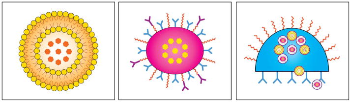
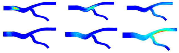
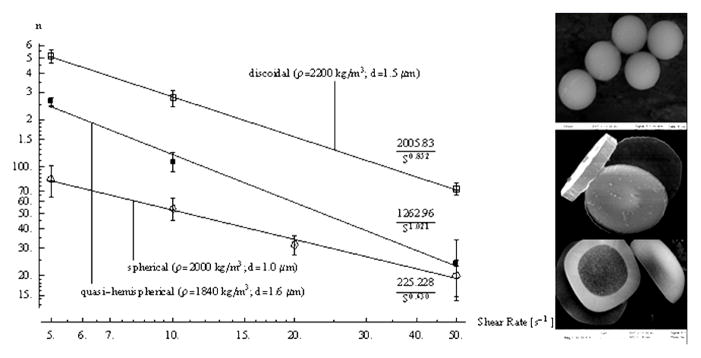
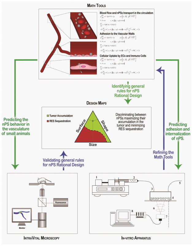
The use of nanoparticles for the early detection, cure, and imaging of diseases has been proved already to have a colossal potential in different biomedical fields, such as oncology and cardiology. A broad spectrum of nanoparticles are currently under development, exhibiting differences in (i) size, ranging from few tens of nanometers to few microns; (ii) shape, from the classical spherical beads to discoidal, hemispherical, cylindrical, and conical; (iii) surface functionalization, with a wide range of electrostatic charges and biomolecule conjugations. Clearly, the library of nanoparticles generated by combining all possible sizes, shapes, and surface physicochemical properties is enormous. With such a complex scenario, an integrated approach is here proposed and described for the rational design of nanoparticle systems (nanovectors) for the intravascular delivery of therapeutic and imaging contrast agents. The proposed integrated approach combines multiscale/multiphysics mathematical models with in vitro assays and in vivo intravital microscopy (IVM) experiments and aims at identifying the optimal combination of size, shape, and surface properties that maximize the nanovectors localization within the diseased microvasculature.


Figures

Fig. 1
Fig. 1
(a) First-generation nanovectors, as the currently clinical liposomes, comprise a container (phospholipidic bilayer in yellow) and an active principle (red dots). They localize in the tumor by enhanced permeation and retention (EPR); (b) Second-generation nanovectors further possess the ability for the targeting of their therapeutic action via antibodies and other biomolecules, remote activation, or responsiveness to environment; (c) Third-generation nanovectors such as multistage agents are capable of more complex functions, such a time-controlled deployment of multiple waves of active nanoparticles, deployed across different biological barriers and with different sub-cellular targets.
Fig. 2
Fig. 2
Concentration C of nanovectors transported along an authentically complex vascular network (top) and wall concentration Cw (bottom) of nanovectors at t=3, 6 and 9 sec after a bolus injection within the inlet section (from direction from left to right).
Fig. 3
Fig. 3
The dynamics of an ellipsoidal particle (aspect ratio 0.5) moving in proximity to a vessel wall. The particle is drifting towards the wall at the bottom of the plot [adapted from Lee et al., 2009].
Fig. 4
Fig. 4
The probability of adhesion for a spheroidal particle in a capillary flow [adapted from Decuzzi et al., 2004]
Fig. 5
Fig. 5
Number of marginating nanoparticles as a function of the shear rate and particle shape [adapted from Decuzzi and Ferrari, 2006]
Fig. 6
Fig. 6
Characteristic half-time τw for the receptor mediated uptake of ellipsoidal particles with aspect ratio Γ [adapted from Decuzzi and Ferrari, 2009].
Fig. 7
Fig. 7
Design maps for spherical beads [adapted from Decuzzi and Ferrari, 2008]
Fig. 8
Fig. 8
The experimental apparatus: 1. syringe (and pump); 2. nanovectors in solution, 3 flow chamber; 4. cell culture dish; 5. gasket; 6. microscope; 7. acquisition system; 8 PC
Fig. 9
Fig. 9
From left: Bright field and fluorescence images of 1 μm fluorescent particles adhering to a sub-confluent layer of Human Umbilical Vein endothelial Cells (HUVECs); fluorescent image; schematic for image analysis; number of adhering particles over time (Decuzzi et al., 2007).
Fig. 10
Fig. 10
Intravital microscopy examples. A) Brightfiled imaging of mouse cremaster showing three rolling leukocytes (arrows) in a convergent venule. B) Brightfield imaging of rat mesentery with a convergent venule (ven.) and arteriole (art.). C) Epifluorescence image of limbal microvessels of the mouse cornea, following intravascular injection of a fluorescently-labeled macromolecule. Bar = 50 μm.
Fig. 11
Fig. 11
Two fluorescently-labeled microparticles (1 μm diameter, arrows) in a mouse cremaster arteriole. Bar = 20 μm
Fig. 12
Fig. 12
The integrated approach and the interaction among the three fundamental components.

Similar articles

Cited by

References

    1. Adams GP, Schier R, McCall AM, Simmons HH, Horak EM, Alpaugh RK, Marks JD, Weiner LM. High affinity restricts the localization and tumor penetration of single-chain fv antibody molecules. Cancer Res. 2001;61:4750. - PubMed
    1. Allen TM. Ligand-targeted therapeutics in anticancer therapy Nat. Rev Drug Discov. 2002;2:750. - PubMed
    1. Allen TM. Ligand-targeted therapeutics in anticancer therapy. Nat Rev Drug Discov. 2002;2:750. - PubMed
    1. Allen TM, Ahmad I, Lopes de Menezes DE, Moase EH. Immunoliposome-mediated targeting of anti-cancer drugs in vivo. Biochem Soc Trans. 1995;23:1073–1079. - PubMed
    1. Allen TM, Sapra P, Moase E, Moreira J, Iden D. Adventures in targeting. J Liposome Res. 2002;12:5–12. - PubMed

Publication types