. 2010 Nov 12;285(46):35706-18.
doi: 10.1074/jbc.M110.125609. Epub 2010 Aug 31.

Regulated secretion of acid sphingomyelinase: implications for selectivity of ceramide formation

Affiliations

Regulated secretion of acid sphingomyelinase: implications for selectivity of ceramide formation

Russell W Jenkins et al. J Biol Chem. .

Abstract

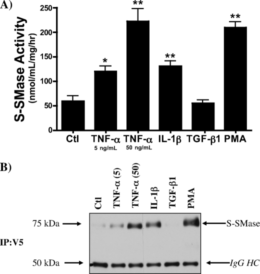
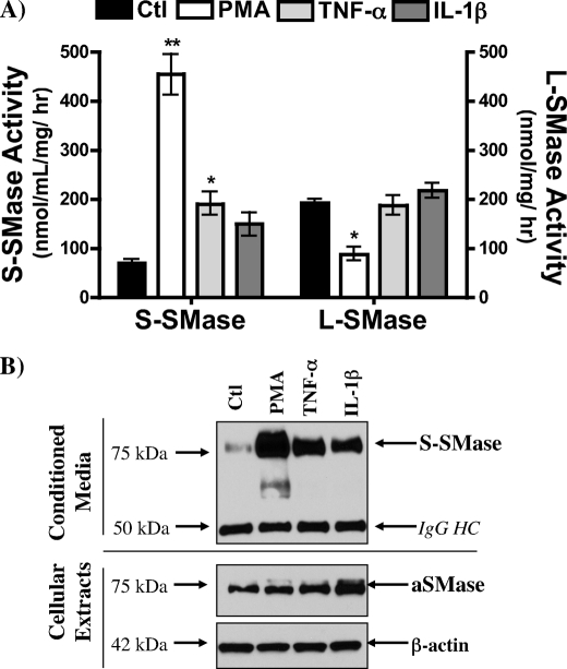
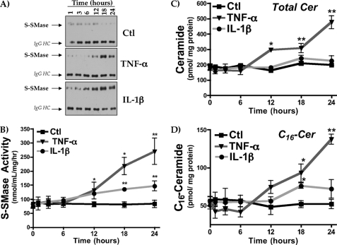
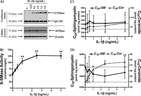
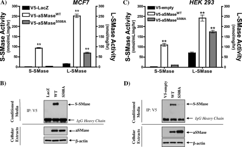
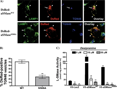
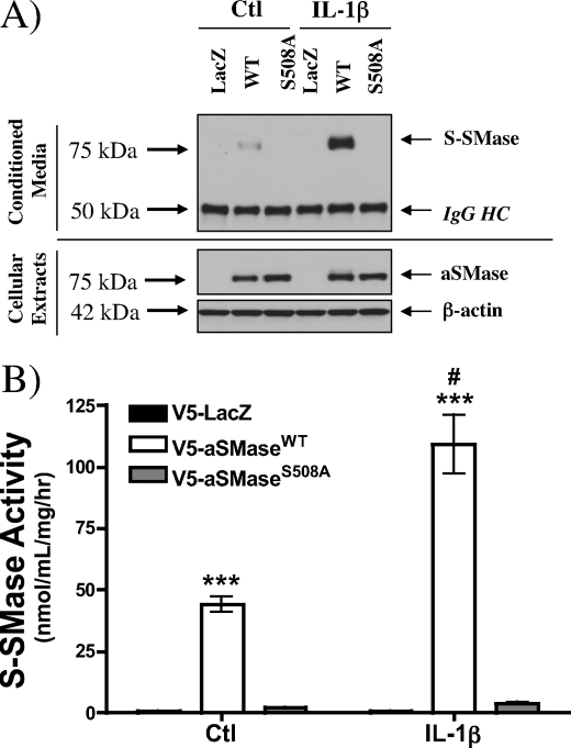
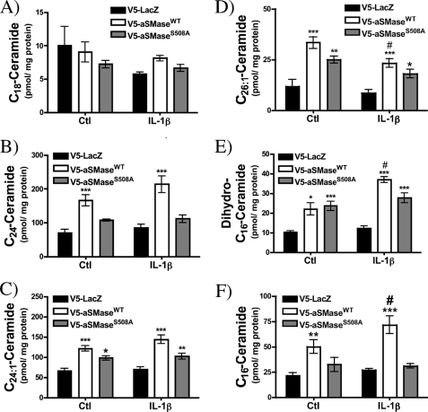
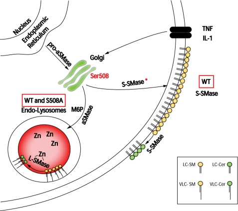
The acid sphingomyelinase (aSMase) gene gives rise to two distinct enzymes, lysosomal sphingomyelinase (L-SMase) and secretory sphingomyelinase (S-SMase), via differential trafficking of a common protein precursor. However, the regulation of S-SMase and its role in cytokine-induced ceramide formation remain ill defined. To determine the role of S-SMase in cellular sphingolipid metabolism, MCF7 breast carcinoma cells stably transfected with V5-aSMase(WT) were treated with inflammatory cytokines. Interleukin-1β and tumor necrosis factor-α induced a time- and dose-dependent increase in S-SMase secretion and activity, coincident with selective elevations in cellular C(16)-ceramide. To establish a role for S-SMase, we utilized a mutant of aSMase (S508A) that is shown to retain L-SMase activity, but is defective in secretion. MCF7 expressing V5-aSMase(WT) exhibited increased S-SMase and L-SMase activity, as well as elevated cellular levels of specific long-chain and very long-chain ceramide species relative to vector control MCF7. Interestingly, elevated levels of only certain very long-chain ceramides were evident in V5-aSMase(S508A) MCF7. Secretion of the S508A mutant was also defective in response to IL-1β, as was the regulated generation of C(16)-ceramide. Taken together, these data support a crucial role for Ser(508) in the regulation of S-SMase secretion, and they suggest distinct metabolic roles for S-SMase and L-SMase.


Figures

FIGURE 1.
FIGURE 1.
Effect of inflammatory cytokines on S-SMase activity and secretion. Serum-starved V5-aSMaseWT MCF7 cells treated with vehicle (PBS), TNF-α (5, 50 ng/ml), IL-1β (2.5 ng/ml), TGF-β1 (2.5 ng/ml), or PMA (100 nm) for 24 h. Conditioned media were collected the following day as described under “Experimental Procedures” and processed for: A, S-SMase activity and B, immunoblotting for V5-S-SMase following immunoprecipitation (n = 4). For S-SMase activity, less than 1% of measured S-SMase activity was due to endogenous S-SMase activity. For V5 immunoblotting, equal volumes of immune complexes (15 μl/lane) were resolved by SDS-PAGE. Data are expressed as mean ± S.E. *, p < 0.05; **, p < 0.01 (one-way ANOVA). IP, immunoprecipitation.
FIGURE 2.
FIGURE 2.
Effect of inflammatory cytokines on S-SMase and L-SMase activity. After overnight serum starvation, MCF7 V5-aSMaseWT cells were treated with vehicle (PBS), PMA (100 nm), TNF-α (50 ng/ml), or IL-1β (2.5 ng/ml) for 18 h. Conditioned media and adherent cells were then collected as described under “Experimental Procedures” and processed for: A, S-SMase and L-SMase activity and B, immunoblotting for V5-S-SMase, V5-aSMase, and β-actin (n = 4). Equal volumes of immune complexes (15 μl/lane) and cellular lysates (10 μg of protein/lane) were resolved by SDS-PAGE for V5 and β-actin immunoblotting. Additional processing of S-SMase may also occur in response to stimulation with inflammatory stimuli, as a 55–60-kDa V5-immunoreactive fragment was detected (B, second lane, top). This band was also evident in IL-1β- and TNF-α-treated cells upon prolonged exposure (data not shown). Data are expressed as mean ± S.E. *, p < 0.05; **, p < 0.01 (one-way ANOVA, Dunnett's post-test).
FIGURE 3.
FIGURE 3.
Effect of inflammatory stimuli on SMPD1 mRNA levels in MCF7 V5-aSMaseWT. After overnight serum starvation, V5-aSMaseWT MCF7 cells were treated with vehicle (PBS), PMA (100 nm), or IL-1β (2.5 ng/ml) for 18 h. RNA was extracted, converted to cDNA, and mean normalized expression (MNE) was determined for aSMase (SMPD1) relative to β-actin as described under “Experimental Procedures.” Data are expressed as mean ± S.E. (n = 4). **, p < 0.01 (one-way ANOVA, Dunnett's post-test).
FIGURE 4.
FIGURE 4.
Protein stability of S-SMase and L-SMase. V5-aSMaseWT MCF7 cells were treated with vehicle (PBS), PMA (100 nm), or IL-1β (2.5 ng/ml) for 18 h. Adherent cells (A and B) and conditioned media (C and D) were then collected as described under “Experimental Procedures” either immediately (0 h) or after 6, 12, or 24 h of cycloheximide treatment (CHX, 50 μg/ml in PBS). Cellular extracts were assessed for: A, L-SMase activity and B, V5-aSMase protein levels by immunoblotting with β-actin as a loading control. Conditioned media were analyzed for: C, S-SMase activity and D, V5-S-SMase protein levels by immunoblotting following immunoprecipitation by V5 mAb. Equal volumes of immune complexes (15 μl/lane) and cellular lysates (10 μg of protein/lane) were resolved by SDS-PAGE for V5 and β-actin immunoblotting. L-SMase activity (nmol/mg/h) and S-SMase activity (nmol/ml/h) were determined for each sample and expressed as percent of 0 h. CHX time point for each treatment group are shown: control (Ctl), IL-1β, and PMA (n = 3). Data are expressed as mean ± S.E. *, p < 0.05 versus control group; #, p < 0.05 versus 0 h. CHX (two-way ANOVA, Bonferroni post test).
FIGURE 5.
FIGURE 5.
Kinetics of S-SMase secretion and ceramide generation in response to IL-1β and TNF-α. 3 × 10⁵ were seeded in 60-mm dishes. After overnight serum starvation, V5-aSMaseWT MCF7 cells were treated with PBS (control, Ctl), IL-1β (2.5 ng/ml), and TNF-α (50 ng/ml) for the indicated amounts of time and conditioned media were collected and assessed for S-SMase secretion by: A, V5 immunoprecipitation and B, by S-SMase activity. Equal volumes of immune complexes (15 μl/lane) were resolved by SDS-PAGE for V5 immunoblotting. Cellular extracts were prepared as described above and submitted for sphingolipidomic analysis for total cellular Cer (C). The levels of a particular Cer species, (D) C16-Cer, are displayed (n ≥ 3). Data are expressed as mean ± S.E. *, p < 0.05; **, p < 0.01 (one-way ANOVA, Dunnett's post test for each group compared with the 0-h time point).
FIGURE 6.
FIGURE 6.
Dose-response of S-SMase secretion and ceramide generation in response to IL-1β. IL-1β was administered to V5-aSMaseWT MCF7 at the indicated doses for 18 h and conditioned media were collected and assessed for S-SMase secretion by: A, V5 immunoprecipitation and B, by S-SMase activity. Cellular levels of V5-aSMase were assessed by Western blotting with β-actin as a loading control. Cellular extracts were prepared as described above and submitted for sphingolipidomic analysis. Equal volumes of immune complexes (15 μl/lane) and cellular lysates (10 μg of protein/lane) were resolved by SDS-PAGE for V5 and β-actin immunoblotting. Levels of (C) C24-SM and C24-Cer and (D) C16-SM and C16-Cer are displayed (n = 4). Data are expressed as mean ± S.E. *, p < 0.05; **, p < 0.01 (one-way ANOVA, Dunnett's post test).
FIGURE 7.
FIGURE 7.
Mutation of Ser508 to Ala abolishes constitutive secretion of S-SMase. After overnight serum starvation, conditioned medium (S-SMase) and adherent cells (L-SMase) were collected for analysis from MCF7 stables (A and B) and HEK stables (C and D) as described under “Experimental Procedures.” V5-S-SMase was concentrated from conditioned medium by immunoprecipitation (IP). Equal volumes of immune complexes (15 μl/lane) and cellular lysates (10 μg of protein/lane) were resolved by SDS-PAGE for V5 and β-actin immunoblotting. Data are expressed as mean ± S.E. (n ≥ 3). *, p < 0.05; **, p < 0.01 (one-way ANOVA, Dunnett's post test).
FIGURE 8.
FIGURE 8.
Lysosomal targeting of S508A mutant. A, HEK 293 stably expressing DsRed-aSMaseWT and DsRed-aSMaseS508A were fixed and processed for immunostaining with anti-TGN46 and anti-LAMP1 antibodies. Small arrows denote areas of LAMP1+DsRed+ structures, whereas long arrows denote TGN46+DsRed+ structures. B, colocalization of DsRed aSMase (WT and S508A) with TGN46 was quantified as percent of DsRed-positive structures that colocalized with TGN46 vesicles. Data are expressed as mean ± S.E. from three independent experiments (**, p < 0.01, unpaired Student's t test). C, V5-LacZ, V5-aSMaseWT, and V5-aSMaseS508A MCF7 seeded in 60-mm dishes were serum starved overnight prior to treatment with PBS or the indicated dose of desipramine for 2 h. Lysates prepared from adherent cells were assayed for L-SMase activity as described above (20 μg of lysate, 30 min at 37 °C). Data are expressed as mean ± S.E. (n = 3). *, p < 0.05; **, p < 0.01 (one-way ANOVA, Dunnett's post test).
FIGURE 9.
FIGURE 9.
Effect of IL-1β on S-SMase secretion in MCF7 stable cell lines. Conditioned media and cellular extracts were prepared from V5-LacZ, V5-aSMaseWT, and V5-aSMaseS508A MCF7 following 18 h of PBS and IL-1β (2.5 ng/ml). Conditioned media were collected and assessed for S-SMase secretion by: A, V5 immunoprecipitation and B, by S-SMase activity. For cellular V5-aSMase, membranes were probed with V5 and β-actin as a loading control. Equal volumes of immune complexes (15 μl/lane) and cellular lysates (10 μg of protein/lane) were resolved by SDS-PAGE for V5 and β-actin immunoblotting. Data are expressed as mean ± S.E. (n ≥ 3). *, p < 0.05; **, p < 0.01; ***, p < 0.001 versus LacZ; #, p < 0.05 versus untreated control (two-way ANOVA, Bonferroni post test).
FIGURE 10.
FIGURE 10.
Effect of IL-1β on the cellular ceramide profile in MCF7 stable cell lines. Cellular extracts were prepared from V5-LacZ, V5-aSMaseWT, and V5-aSMaseS508A MCF7 following 18 h of PBS and IL-1β (2.5 ng/ml). Shown are representative Cer species that are: A, unaffected by WT or S508A aSMase and unchanged by IL-1β (e.g. C18-Cer); B, affected by WT, but not S508A aSMase, and unchanged by IL-1β (e.g. C24-Cer); C, affected by WT and S508A aSMase, but unchanged by IL-1β (e.g. C24:1-Cer); D, affected by WT and S508A aSMase, and decreased by IL-1β (e.g. C26:1-Cer); E, affected by WT and S508A aSMase, and increased by IL-1β (e.g. dihydro-C16-Cer); and F, affected by WT, but not S508A aSMase, and increased by IL-1β (e.g. C16-Cer). Data are expressed as mean ± S.E. (n ≥ 3). *, p < 0.05; **, p < 0.01; ***, p < 0.001 versus LacZ; #, p < 0.05 versus untreated control (Ctl) (two-way ANOVA, Bonferroni post test).
FIGURE 11.
FIGURE 11.
Schematic representation of S-SMase and L-SMase localization and metabolic action. In response to inflammatory cytokine challenge, the common aSMase precursor (pro-aSMase) is selectively shuttled away from the lysosomal pathway and toward the Golgi secretory pathway. Up-regulation of S-SMase in response to TNF-α or IL-1β is associated with a selective elevation in particular the long-chain Cer species (LC-Cer) (e.g. C16-Cer), which is not evident in cells overexpressing the secretion-incompetent S508A mutant. On the other hand, both WT and S508A aSMase exhibit elevations in L-SMase activity with associated increases in selective very long-chain Cer species (VLC-Cer) (e.g. C26:1-Cer). Ser508 is a putative phosphorylation site that may influence trafficking of pro-aSMase at the level of the Golgi.

Similar articles

Cited by

References

    1. Hannun Y. A., Obeid L. M. (2008) Nat. Rev. Mol. Cell Biol. 9, 139–150 - PubMed
    1. Gulbins E., Li P. L. (2006) Am. J. Physiol. Regul. Integr. Comp. Physiol. 290, R11–26 - PubMed
    1. Pettus B. J., Chalfant C. E., Hannun Y. A. (2004) Curr. Mol. Med. 4, 405–418 - PubMed
    1. Ogretmen B., Hannun Y. A. (2004) Nat. Rev. Cancer. 4, 604–616 - PubMed
    1. Goñi F. M., Alonso A. (2002) FEBS Lett. 531, 38–46 - PubMed

Publication types

MeSH terms

LinkOut - more resources