. 2010 Nov 5;285(45):34390-400.
doi: 10.1074/jbc.M110.120576. Epub 2010 Aug 31.

Identification of an endothelin-converting enzyme-2-specific fluorigenic substrate and development of an in vitro and ex vivo enzymatic assay

Affiliations

Identification of an endothelin-converting enzyme-2-specific fluorigenic substrate and development of an in vitro and ex vivo enzymatic assay

Tanja Ouimet et al. J Biol Chem. .

Abstract

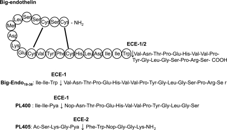
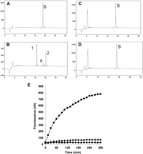
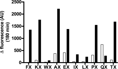
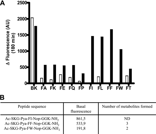
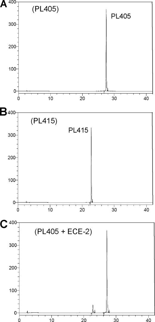
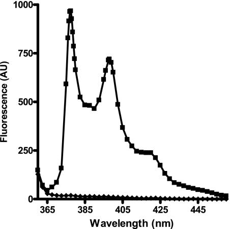
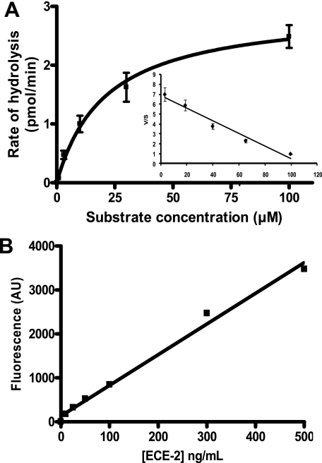
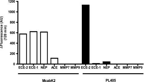
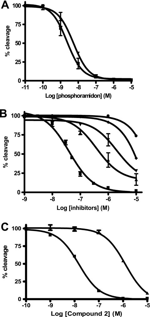
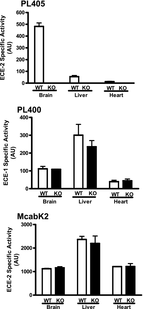
Endothelin-converting enzyme-2 (ECE-2) is a membrane-bound zinc-dependent metalloprotease that shares a high degree of sequence homology with ECE-1, but displays an acidic pH optimum characteristic of maturing enzymes acting late in the secretory pathway. Although ECE-2, like ECE-1, can cleave the big endothelin intermediate to produce the vasoconstrictive endothelin peptide, its true physiological function remains to be elucidated, a task that is hampered by the lack of specific tools to study and discriminate ECE-2 from ECE-1, i.e. specific substrates and/or specific inhibitors. To fill this gap, we searched for novel ECE-specific peptide substrates. To this end, peptides derived from the big endothelin intermediate were tested using ECE-1 and ECE-2, leading to the identification of an ECE-1-specific substrate. Moreover, screening of our proprietary fluorigenic peptide Fluofast® libraries using ECE-1 and ECE-2 allowed the identification of Ac-SKG-Pya-F-W-Nop-GGK-NH(2) (PL405), as a specific and high affinity ECE-2 substrate. Indeed, ECE-2 cleaved PL405 at the Pya-F amide bond with a specificity constant (k(cat)/K(m)) of 8.1 ± 0.9 × 10(3) M(-1) s(-1). Using this novel substrate, we also characterized the first potent (K(i) = 7.7 ± 0.3 nM) and relatively selective ECE-2 inhibitor and developed a quantitative fluorigenic ECE-2 assay. The assay was used to study the ex vivo ECE-2 activity in wild type and ECE-2 knock-out tissues and was found to truly reflect ECE-2 expression patterns. The PL405 assay is thus the first tool to study ECE-2 inhibition using high throughput screening or for ex vivo ECE-2 quantification.


Figures

FIGURE 1.
FIGURE 1.
Top sequence, amino acid sequence of the native big endothelin 1–38 intermediate containing the cyclic 21-amino acid sequence of endothelin 1, which is released after cleavage of the Trp21–Val22 bond by ECE-1 and ECE-2. Second sequence, amino acid sequence of the synthetic big endothelin 19–38 peptide containing the Trp-Val scissile bond in positions 3 and 4 of the peptide, which is cleaved by ECE-1 only. Third sequence, amino acid sequence of the selective ECE-1 peptide substrate, PL400 (19), based on the big endothelin 19–35, in which the scissile bond is formed by the fluorophore and repressor pair Pya and Nop. Fourth sequence, the PL405 peptide substrate cleaved exclusively by ECE-2 at the Pya-Phe amide bond.
FIGURE 2.
FIGURE 2.
Cleavage of big endothelin 19–38 (human) and of PL400 by ECE-1 and ECE-2. A–D, big endothelin 19–38 (S, 50 μm) was incubated for 3 h at 37 °C either in 100 mm HEPES pH 6.8 buffer alone (A) or in buffer with 1 μg/ml ECE-1 (B), and the products were separated by HPLC on a Kromasil C18 column (5 μm, 100 Å, 4.6 × 150 mm). The same experiment was performed with ECE-2 by incubating the big endothelin 19–38 peptide (50 μm) alone in 200 mm sodium acetate, pH 5.5, for 3 h at 37 °C (C) or in buffer with 1 μg/ml of ECE-2 (D). The reaction products were separated according to a 0–60% gradient of solvent B in 30 min. Solvent A was 0.1% trifluoroacetic acid in H2O. Solvent B was 0.1% trifluoroacetic acid in CH3CN. The flow rate was 1 ml/min. Absorbance detection (UV) was 210 nm. Hydrolysis products 1 and 2 were found to correspond to big endothelin 22–38 and to the Ile-Ile-Trp tri-peptide, respectively. E, the PL400 peptide (20 μm) was incubated for 5 h at 37 °C either alone in ECE-1 reaction buffer (♦) or in the same buffer with 1 μg/ml of ECE-1 (■). For ECE-2, 20 μm PL400 was incubated either in ECE-2 reaction buffer (●) or in buffer with 1 μg/ml of ECE-2 (▴).
FIGURE 3.
FIGURE 3.
Comparative screening of the Ac-SKG-Pya-N1-X-Nop-GGK-CONH2 peptide libraries. Either ECE-1 (white) or ECE-2 (black) at 1 μg/ml was incubated at 37 °C for 3 h with 100 μm of each peptide library in a final volume of 100 μl of 100 mm HEPES, pH 6.8, or 200 mm sodium acetate, pH 5.5, respectively. Control base fluorescence was measured by incubating the peptide library alone in 100 μl of its reaction buffer. The histogram shows the deltas of fluorescence, which correspond to the fluorescence of the assay minus that of the base fluorescence of the library at 180 min.
FIGURE 4.
FIGURE 4.
A, comparative ECE-1 and ECE-2 enzymatic activities of the deconvoluted Ac-SKG-Pya-FX-Nop-GGK-CONH2 peptide library. Either ECE-1 (white) or ECE-2 (black) at 1 μg/ml was incubated for 3 h at 37 °C with each peptide (10 μm) in a final volume of 100 μl of 100 mm HEPES, pH 6.8, or 200 mm sodium acetate, pH 5.5, respectively. Positive control incubation was performed in parallel using the McabK2 substrate (BK) at 10 μm with both enzymes, in the same conditions. B, peptide sequence of the three best peptide substrates. The base fluorescence of these three peptides (10 μm) in the ECE-2 reaction buffer was measured. The number of hydrolysis products was observed by HPLC after incubation with 1 μg/ml of ECE-2, and they were subsequently analyzed by mass spectrometry.
FIGURE 5.
FIGURE 5.
HPLC analysis of the synthetic substrate PL405 cleavage by ECE-2. The HPLC conditions were Kromasil C18 column (5 μm, 100 Å, 4.6 × 150 mm); gradient 10–90% solvent B in 60 min. Solvent A was 0.1% trifluoroacetic acid in H2O. Solvent B was 0.1% trifluoroacetic acid in CH3CN. The flow rate was 1 ml/min. Absorbance detection (UV) was 343 nm. A, injection of 100 μl of 10 μm of PL405 substrate in 200 mm sodium acetate, pH 5.5, 0.2 n HCl. The peak of the substrate has a retention time of 27.8 min. B, injection of 100 μl of 10 μm of Ac-SKG-Pya cleavage product PL415 in the same buffer. The retention time of the peak was 23.1 min. C, the PL405 substrate (20 μm) was incubated 3 h at 37 °C in 200 mm sodium acetate pH 5.5 with 500 ng/ml of ECE-2. The reaction was stopped with 0.2 n HCl, and 100 μl of the reaction were separated by HPLC.
FIGURE 6.
FIGURE 6.
Emission spectra (λex = 343 nm) of 1 μm of the PL405 substrate (♦) and of 1 μm of the PL415 cleavage product (■) in 200 mm sodium acetate, pH 5.5.
FIGURE 7.
FIGURE 7.
A, kinetic parameters of the ECE-2 enzymatic activity using PL405. Increasing concentrations of PL405 were incubated 60 min at 37 °C in 200 mm sodium acetate, pH 5.5, containing 250 ng/ml of ECE-2. The mean values of two independent experiments performed in duplicate are represented. The Km of the substrate is 21 ± 4 μm, the catalytic constant (kcat) is 0.17 ± 0.02 s−1, and the kcat/Km is 8.1 × 10³ ± 0.9 × 10³ m−1·s−1. B, cleavage of the PL405 (20 μm) by ECE-2 as a function of enzyme concentrations in vitro (incubation 3 h at 37 °C).
FIGURE 8.
FIGURE 8.
McabK2versus PL405 enzymatic assay: specificity of the PL405 substrate. Histogram showing the specific fluorescent signal measured (total fluorescence minus fluorescence of substrate alone in buffer) with either substrate in the presence of 250 ng/ml of ECE-2, ECE-1, NEP, ACE, MMP7, or MMP9 after a 120-min incubation at 37 °C in 200 mm sodium acetate, pH 5.5. In these conditions, whereas McabK2 is cleaved by ECE-1 and NEP as efficiently as by ECE-2, the novel PL405 substrate is exclusively metabolized by ECE-2.
FIGURE 9.
FIGURE 9.
A, inhibition of ECE-2 enzymatic activity by phosphoramidon using either McabK2 (▾) or PL405 (■) fluorogenic substrates. Increasing concentrations of phosphoramidon were incubated 10 min at 37 °C with 100 ng/ml of ECE-2 in 200 mm sodium acetate, pH 5.5. The reactions were initiated by the addition of either 10 μm McabK2 or 20 μm PL405 followed by a 30- or 60-min incubation at 37 °C, respectively. B, inhibition of ECE-2 by various metalloprotease inhibitory compounds. Different concentrations of either compound 1 (■), kelatorphan (▴), retrothiorphan (▾), thiorphan (♦), or captopril (●) were incubated 10 min at 37 °C with 100 ng/ml of ECE-2 in 200 mm sodium acetate, pH 5.5. The reactions were initiated by the addition of 20 μm PL405 followed by a 60-min incubation at 37 °C. C, inhibitory potency of compound 2 against ECE-2 using the PL405 assay (■) compared with that against ECE-1 using the McabK2 substrate (▴), revealing a discriminating factor of 62. The obtained Ki values from the conversion of the obtained IC50 values are reported in Table 1.
FIGURE 10.
FIGURE 10.
Ex vivo ECE-2 enzymatic activity in WT and ECE-2 knock-out (KO) tissues. The specific ECE-2 enzymatic activity in solubilized membrane preparations from brain, liver, and heart tissue from either WT or KO animals was measured using 20 μm of the specific PL405 substrate (top panel). The same preparations were used to monitor specific ECE-1 activity (middle panel) using 100 μm PL400. The bottom panel shows the nondiscriminative results obtained using the McabK2 substrate (10 μm). The enzymatic reaction was monitored for 5 h at 37 °C. The results depicted are the means from two independent experiments performed using tissues from two WT and two KO animals.

Similar articles

Cited by

References

    1. Xu D., Emoto N., Giaid A., Slaughter C., Kaw S., deWit D., Yanagisawa M. (1994) Cell 78, 473–485 - PubMed
    1. Emoto N., Yanagisawa M. (1995) J. Biol. Chem. 270, 15262–15268 - PubMed
    1. Roques B. P., Noble F., Daugé V., Fournié-Zaluski M. C., Beaumont A. (1993) Pharmacol Rev. 45, 87–146 - PubMed
    1. Valdenaire O., Richards J. G., Faull R. L., Schweizer A. (1999) Brain Res. Mol. Brain Res. 64, 211–221 - PubMed
    1. Kiryu-Seo S., Sasaki M., Yokohama H., Nakagomi S., Hirayama T., Aoki S., Wada K., Kiyama H. (2000) Proc. Natl. Acad. Sci. U.S.A. 97, 4345–4350 - PMC - PubMed

MeSH terms

LinkOut - more resources