. 2010 Nov 1;588(Pt 21):4117-29.
doi: 10.1113/jphysiol.2010.195099. Epub 2010 Aug 31.

Orexin neurons are indispensable for stress-induced thermogenesis in mice

Affiliations

Orexin neurons are indispensable for stress-induced thermogenesis in mice

Wei Zhang et al. J Physiol. .

Abstract

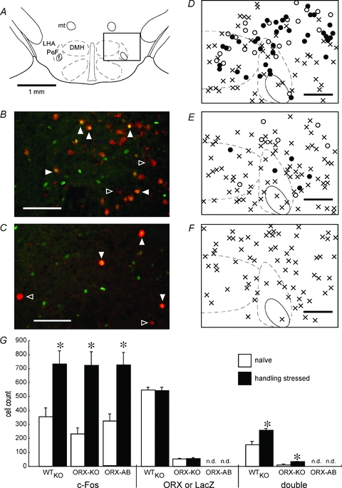
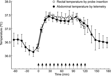
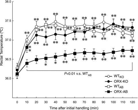
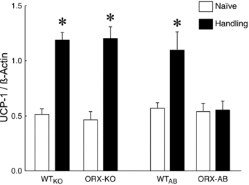
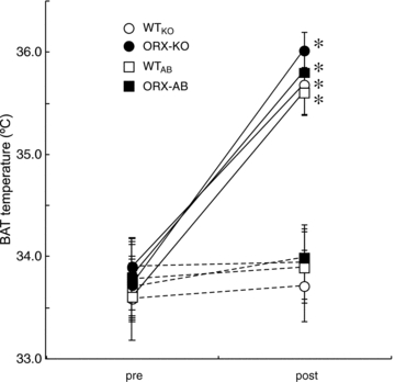
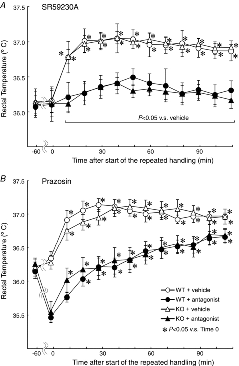
Orexin neurons contribute to cardiovascular, respiratory and analgesic components of the fight-or-flight response against stressors. Here, we examined whether the same is true for stress-induced hyperthermia. We used prepro-orexin knockout mice (ORX-KO) and orexin neuron-ablated mice (ORX-AB) in which the latter lack not only orexin, but also other putative neurotransmitter/modulators contained in the orexin neurons. In response to repetitive insertion of a temperature probe into their rectum (handling stress), ORX-KO mice showed a normal temperature change as compared to that of wild-type littermates (WT) while ORX-AB showed an attenuated response. Stress-induced expression of uncoupling protein-1, a key molecule in non-shivering thermogenesis in the brown adipose tissue (BAT), was also blunted in ORX-AB but not in ORX-KO. When the BAT was directly activated by a β3 adrenergic agonist, there was no difference in the resultant BAT temperature among the groups, indicating that BAT per se was normal in ORX-AB. In WT and ORX-KO, handling stress activated orexin neurons (as revealed by increased expression of c-Fos) and the resultant hyperthermia was largely blunted by pre-treatment with a β3 antagonist. This observation further supports the notion that attenuated stress-induced hyperthermia in ORX-AB mice was caused by a loss of orexin neurons and abnormal BAT regulation. This study pointed out, for the first time, the possible importance of co-existent neurotransmitter/modulators in the orexin neurons for stress-induced hyperthermia and the importance of integrity of the orexin neurons for full expression of multiple facets of the fight-or-flight response.


Figures

Figure 5
Figure 5. Immunohistochemical evidence for activation of orexin neurons by handling stress
A, schematic drawing of a coronal section of the mouse brain showing structure of the hypothalamus. A rectangle denotes examined area (711 × 944 μm). Both sides were examined in the actual experiment although only a right side window was depicted for simplicity. DMH, dorsomedial hypothalamus; f, fornix; LHA, lateral hypothalamic area; mt, mammillothalamic tract; PeF, perifornical area. B, representative photograph of double immunostaining for orexin and c-Fos in the hypothalamus of a wild-type (WT) mouse sampled after 2 h of repetitive handling stress. Orexin was stained in red and c-Fos was stained in green. Yellow designates cells stained for both orexin and c-Fos. Filled triangles indicate double-stained cells and open triangles indicate orexin-containing but not c-Fos-expressing cells. C, representative photograph of double immunostaining for LacZ (β-galactosidase) and c-Fos in the hypothalamus of an orexin knockout (ORX-KO) mouse sampled after 2 h of repetitive handling stress. LacZ gene was introduced into the ORX-KO mice instead of the normal orexin gene. LacZ was stained in red and c-Fos was stained in green. Yellow designates cells stained for both LacZ and c-Fos. Filled triangles indicate double-stained cells and open triangles indicate LacZ-containing but not c-Fos expressing cells. Bar, 100 μm in B and C. D, typical distribution of double-stained cells (filled circle), orexin-positive but not c-Fos positive cells (open circle), and c-Fos-positive but not orexin-positive cells (×) in the examination window of a WT mouse. E, typical distribution of double-stained cells (filled circle), LacZ-positive but not c-Fos-positive cells (open circle), and c-Fos positive but not LacZ-positive cells (×) in the examination window of a ORX-KO mouse. F, typical distribution of c-Fos-positive cells (×) in the examination window of a orexin neuron-ablated (ORX-AB) mouse. Note that there is no apparent difference in the distribution of c-Fos-positive cells among D, E and F. Bar, 200 μm in D–F. G, numbers of c-Fos-, orexin (ORX)- and LacZ-immunopositive cells and double-stained cells (c-Fos and ORX in WT or in ORX-AB, and c-Fos and LacZ in ORX-KO) in the hypothalamus. Every 4th section in an animal (6 sections per mouse) was examined. Data are presented as mean ±s.e.m. of 4 mice in a group except for handling-stressed ORX-KO mice (n = 3). *P < 0.05 vs. naïve. No orexin-immunopositive cell was detected (n.d.) in ORX-AB mice.
Figure 1
Figure 1. Comparison between body temperature measurement by rectal temperature probe insertion and abdominal telemetry in wild-type mice
To confirm that the temperature measurement by a rectal probe accurately reflects the body temperature just before probe insertion but subsequently triggers hyperthermia, rectal temperature was repetitively measured at 10 min intervals while abdominal temperature was continuously measured using radio-telemetry. Data are mean ±s.e.m. of 6 wild-type mice. Arrows indicate the timing of the insertion of temperature probe into the animal's rectum.
Figure 2
Figure 2. Effect of repeated handling stress on rectal temperature of mice of four genotypes
The temperature measurement (insertion of thermistor probe into the animal's rectum) itself was used as a stressor and repeatedly applied at 10 min intervals for 2 h. Data are presented as mean ±s.e.m. of orexin knockout mice (ORX-KO, n = 37), orexin neuron-ablated mice (ORX-AB, n = 32) and their corresponding wild-type littermates (WTKO, n = 22 and WTAB, n = 21). *P < 0.05, **P < 0.01 compared with baseline value at time 0 (Dunnett's post hoc test).
Figure 3
Figure 3. Expression of uncoupling protein (UCP)-1 in the brown adipose tissue from stressed or naïve mice of four genotypes
Brown adipose tissue (BAT) was dissected from orexin knockout mice (ORX-KO), orexin neuron-ablated mice (ORX-AB) and their corresponding wild-type littermates (WTKO and WTAB) after handling stress (4 rectal temperature measurements at 10 min intervals) and from naïve (unstressed) mice of the same genotypes. Total RNA was extracted from the BAT and cDNA was reverse transcribed. UCP-1 mRNA was determined by quantitative real-time PCR in triplicate and normalized with β-actin mRNA. Data are presented as mean ±s.e.m. of 7–9 animals. *P < 0.05 vs. naïve. Note that handling stress increased expression of UCP-1 in wild-type and ORX-KO mice but not in ORX-AB mice.
Figure 4
Figure 4. Brown adipose tissue (BAT) function test
Change of BAT temperature in response to a β3-agonist, CL316243, was examined in chloralose- (75 mg kg−1, i.p.) and urethane- (750 mg kg−1, i.p.) anaesthetized mice. Orexin knockout mice (ORX-KO), orexin neuron-ablated mice (ORX-AB) and their corresponding wild-type litter mates (WTKO and WTAB) received i.p. injection of saline (dashed line) or CL316243 (1 mg kg−1, continuous line). Data are presented as mean ±s.e.m. of the peak values during the observation period of 120 min (n = 5 for each group). *P < 0.05 vs. pre-treatment value.
Figure 6
Figure 6. Effect of a β3-antagonist (SR59230A) and an α1-antagonist (prazosin) on handling stress-induced hyperthermia
SR59230A (5 mg kg−1) and prazosin (1 mg kg−1) was intraperitoneally administered 1 h before the start (at time 0) of the handling stress procedure. Thereafter, temperature measurement was repeated 12 times with an interval of 10 min. Open circles indicate vehicle (saline for SR59230A and 3.3% polyethylene glycol in saline for prazosin)-treated wild-type (WT) mice, filled circles indicate antagonist (SR59230A in A and prazosin in B)-treated WT mice. Open triangles indicate vehicle-treated orexin knockout (KO) mice and filled triangles indicate antagonist-treated KO mice. Each point represents mean ±s.e.m. from 8–10 animals. *P < 0.05 compared with baseline value at time 0 (Dunnett's post hoc test). In A, initial rectal temperature was not different between pre-treatment with SR59230A and saline while, in B, initial rectal temperature was significantly lower in the prazosin-treated group than in the vehicle-treated group. Note that SR59230A prevented handling stress-induced hyperthermia in both WT and KO mice whereas prazosin did not or even exaggerated hyperthermia at the later observation periods (see also Results section).

Comment in

  • The peptide or the neuron?
    McAllen R. McAllen R. J Physiol. 2010 Nov 1;588(Pt 21):4067-8. doi: 10.1113/jphysiol.2010.199745. J Physiol. 2010. PMID: 21037314 Free PMC article. No abstract available.

Similar articles

Cited by

References

    1. Abrahamson EE, Leak RK, Moore RY. The suprachiasmatic nucleus projects to posterior hypothalamic arousal systems. Neuroreport. 2001;12:435–440. - PubMed
    1. Arrigoni E, Mochizuki T, Scammell TE. Activation of the basal forebrain by the orexin/hypocretin neurons. Acta Physiol (Oxf) 2010;198:223–235. - PMC - PubMed
    1. Berthoud H-R, Patterson LM, Sutton GM, Morrison C, Zheng H. Orexin inputs to caudal raphé neurons involved in thermal, cardiovascular, and gastrointestinal regulation. Histochem Cell Biol. 2005;123:147–156. - PubMed
    1. Bouwknecht JA, Olivier B, Paylor RE. The stress-induced hyperthermia paradigm as a physiological animal model for anxiety: a review of pharmacological and genetic studies in the mouse. Neurosci Biobehav Rev. 2007;31:41–59. - PubMed
    1. Cannon B, Nedergaard J. Brown adipose tissue: function and physiological significance. Physiol Rev. 2004;84:277–359. - PubMed

Publication types

MeSH terms

LinkOut - more resources