. 2010 Oct 1;123(Pt 19):3226-34.
doi: 10.1242/jcs.072876. Epub 2010 Aug 31.

The exocytic gene secA is required for Dictyostelium cell motility and osmoregulation

Affiliations

The exocytic gene secA is required for Dictyostelium cell motility and osmoregulation

Roberto Zanchi et al. J Cell Sci. .

Abstract

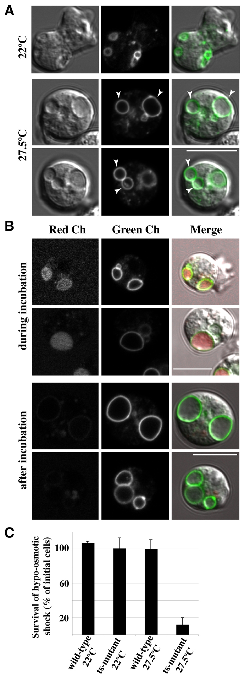
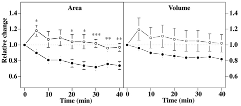
We investigated the link between cell movement and plasma membrane recycling using a fast-acting, temperature-sensitive mutant of the Dictyostelium SecA exocytic protein. Strikingly, most mutant cells become almost paralysed within minutes at the restrictive temperature. However, they can still sense cyclic-AMP (cAMP) gradients and polymerise actin up-gradient, but form only abortive pseudopodia, which cannot expand. They also relay a cAMP signal normally, suggesting that cAMP is released by a non-exocytic mechanism. To investigate why SecA is required for motility, we examined membrane trafficking in the mutant. Plasma membrane circulation is rapidly inhibited at the restrictive temperature and the cells acquire a prominent vesicle. Organelle-specific markers show that this is an undischarged contractile vacuole, and we found the cells are correspondingly osmo-sensitive. Electron microscopy shows that many smaller vesicles, probably originating from the plasma membrane, also accumulate at the restrictive temperature. Consistent with this, the surface area of mutant cells shrinks. We suggest that SecA mutant cells cannot move at the restrictive temperature because their block in exocytosis results in a net uptake of plasma membrane, reducing its area, and so restricting pseudopodial expansion. This demonstrates the importance of proper surface area regulation in cell movement.


Figures

Fig. 1.
Fig. 1.
A temperature-sensitive mutant of SecA. (A) secA is an essential gene. The temperature-sensitive mutant strain HM1325 (ts-mutant) carrying the secA2 allele grows normally at the permissive temperature of 18°C, but not at the restrictive temperature of 27.5°C. A diploid strain harbouring the mutant and wild-type alleles grows normally at both temperatures, showing that the mutant allele is recessive. Cells were picked onto bacterial lawns and photographed after 4 days. Scale bar: 5 mm. (B) Cell morphology at permissive and restrictive temperatures. Mutant and wild type are similar at 18°C. After 30 minutes at 27.5°C, mutant cells become rounded and heavily vacuolated (arrowheads). Freshly starved cells were incubated at 18°C or 27.5°C for 30 minutes before imaging (Normarski). Scale bar: 10 μm. (C) Cell survival at the restrictive temperature. The wild-type (solid line) and temperature-sensitive (HM1325; dashed line) strains were incubated at the restrictive temperature for up to 4 hours (over twice the duration of any assay in this study) and viability determined clonal plating on bacterial lawns at the permissive temperature. No significant change in viability was observed (n=3, error bars represent s.e.m.).
Fig. 2.
Fig. 2.
Random motility of vegetative SecA mutant cells is almost abolished at the restrictive temperature. (A) Tracks of wild-type and SecA mutant cells (HM1325; ts-mutant) at 27.5°C. The tracks of 100 randomly sampled cells are shown centred at the origin. (B) Horizon plot. The fraction of the population moving a given distance from the origin is plotted. More than 75% of mutant cells (lower curve) remain within 2 cell diameters (~ 20 μm) and less than 5% reach a distance of 55 μm from their origin. By contrast, over 50% of the wild-type cells (upper curve) travel at least 85 μm from the origin. (C) Speed distribution. The average mean speed distribution for wild-type cells, white bars, and for the mutant, black bars, is shown. The mean speeds are 8.5±1.6 μm/minute and 1.8±0.4 μm/minute for the wild type and mutant, respectively. Error bars represent s.e.m., n=3; wild type=212 cells, temperature sensitive mutant=348 cells. Cells were washed free of bacteria and plated in axenic medium and incubated for 30 minutes at 27.5°C before imaging for 30 minutes at 3 frames per minute.
Fig. 3.
Fig. 3.
Cells of the SecA mutant can sense chemotactic gradients at the restrictive temperature, but barely move. (A) Tracks of wild-type and SecA mutant cells in a cAMP gradient at the restrictive temperature of 27.5°C. Tracks of aggregation-competent cells in a Dunn chamber (35 wild-type and 45 mutant cells) from paired samples are shown, centred at the origin; filmed for 30 minutes at 3 frames per minute. Representative of four independent experiments; see supplementary material Movie S1. (B) Speed distributions of wild-type and SecA mutant cells. Taken from four independent data sets (124 wild-type cells, white bars; 158 mutant cells, black bars). Mean speeds are wild type, 13.4±1.4 μm/minute; mutant, 2.0±0.3 μm/minute; error bars represent s.e.m. (C) Actin polymerisation produced by uniform cAMP stimulus. All cells display a robust peak 5–10 seconds after stimulation and a more variable second peak about 60 seconds thereafter, which is similar to bulk assays. Aggregation-competent cells were pre-incubated at 27.5°C for 30 minutes and then bath-stimulated with 10 μM cAMP at t0. Actin polymerisation at the cortex of individual cells was monitored using ABD120-GFP as reporter for F-actin, and fluorescence quantified using QuimP. Average from 22 wild-type and 23 mutant cells from three independent experiments. (D) Polarised actin polymerisation induced by a cAMP gradient. Wild-type and mutant cells respond to cAMP gradients by polarised F-actin accumulation up the gradient. This drives expansion of a pseudopod and cell movement in the wild type at either temperature, but the SecA mutant can only extend a pseudopod at the permissive temperature, producing a bulge or even remaining rounded at the restrictive temperature (the extreme cell is from the same population). Cells expressing ABD120-GFP were made aggregation competent and either used immediately or shifted to the restrictive temperature for 30 minutes before imaging. They were stimulated using a needle filled with cAMP (white dot). Scale bar: 10 μm; see supplementary material Movies 2 and 3.
Fig. 4.
Fig. 4.
cAMP relay by aggregation-competent cells. Cells were stimulated with 15 μM 2′-deoxy cAMP and total cAMP produced over 5 minutes (extracellular + cytosolic) and the proportion released determined. (A) SecA mutant (means of five independent experiments; error bars represent s.e.m.). (B) NsfA mutant (means of three independent experiments; error bars represent s.e.m.). No significant differences were observed.
Fig. 5.
Fig. 5.
Contractile vacuole discharge requires SecA activity. (A) The large vesicles formed at the restrictive temperature are undischarged contractile vacuoles. SecA mutant cells, expressing Dajumin-GFP as a contractile vacuole marker, were incubated for 30 minutes at the restrictive temperature of 27.5°C. All large vesicles visible by DIC display the marker. Scale bar: 10 μm. (B) The contractile vacuole ceases discharge at the restrictive temperature. When the contractile vacuole (Dajumin-GFP, green channel) discharges, it becomes accessible to back-filling by small bulk-phase tracers such as Alexa Fluor 594 (red channel). Cells were incubated with dye, either for the first 30 minutes at the restrictive temperature (27.5°C), when it has access to the contractile vacuole (top panels), or for the next 30 minutes, when it does not (lower panels). In both cases, dye in the medium was washed away before observing the cells. Scale bars: 10 μm. (C) The SecA mutant is osmo-sensitive at the restrictive temperature. Cells were transferred from growth medium to water, and after incubation for 30 minutes, their viability was determined by clonal plating on bacterial lawns. Viability of the SecA mutant (ts-mutant 27.5°C) was reduced to less than 10% at the restrictive temperature.
Fig. 6.
Fig. 6.
Ultrastructure of SecA mutant cells. (A) Electron micrographs of representative wild-type and mutant aggregation-competent cells at the restrictive temperature (27.5°C). (a–c) wild-type cell morphology. This is unchanged by the temperature shift. (d–i) Mutant cell morphology at low power, with selected regions at higher power. Two changes in the endomembrane system are apparent: large vacuoles (Va) – undischarged contractile vacuoles – appear, and the number of small vacuoles increases. Often these cluster near the plasma membrane (g and h), sometimes in tight juxtaposition (i). Scale bars: 1 μm (f, for a–f; h, for g,h), 0.2 μm (i). (B) Redistribution of plasma membrane components in the SecA mutant at the restrictive temperature detected using phosphotungstic acid and chromic acid stain. (a–c) Permissive temperature. The plasma membrane stains more strongly than any other membrane and the contractile vacuole does not stain. (d–f) After incubation for 30 minutes at the restrictive temperature. Staining of the plasma membrane is greatly reduced, but a subset of large multi-vesiculated compartments become strongly stained (d and e) and the small vesicles are finely stained, suggesting that they also receive plasma membrane material (f). Scale bars: 1 μm (d, for a and d; c, for b and c; f, for e and f). Aph, autophagosome; MVC, multi-vesciculated compartments; N, nucleus; PM, plasma membrane; Va, vacuole; Ve, vesicle.
Fig. 7.
Fig. 7.
Changes in surface area and volume of mutant cells at the restrictive temperature. Vegetative cells expressing the membrane marker cAR1-GFP were imaged at 27.5°C, taking confocal stacks every 5 minutes. The volume and surface area measurements of wild-type (open circles) and mutant cells (closed circles) are normalised to the initial values (t=0, room temperature). Error bars represent s.e.m. A significant decrease in surface area could be detected in the mutant after 20 minutes at 27.5°C, resulting on average in a ~25% reduction in surface area at 30 minutes. A small decrease in the volume of the mutant is also noticeable, but this is not statistically significant. *P<0.05, **P<0.01, ***P<0.001, two-tailed sample student's t-test (n=6 for the wild-type sample, n=7 for the mutant sample, acquired over 6 different days).

Similar articles

Cited by

References

    1. Aguado-Velasco C., Bretscher M. S. (1999). Circulation of the plasma membrane in Dictyostelium. Mol. Biol. Cell 10, 4419-4427 - PMC - PubMed
    1. Bagorda A., Das S., Rericha E. C., Chen D., Davidson J., Parent C. A. (2009). Real-time measurements of cAMP production in live Dictyostelium cells. J. Cell Sci. 122, 3907-3914 - PMC - PubMed
    1. Betz W. J., Mao F., Smith C. B. (1996). Imaging exocytosis and endocytosis. Curr. Opin. Neurobiol. 6, 365-371 - PubMed
    1. Bloomfield G., Tanaka Y., Skelton J., Ivens A., Kay R. R. (2008). Widespread duplications in the genomes of laboratory stocks of Dictyostelium discoideum. Genome Biol. 9, R75 - PMC - PubMed
    1. Bretscher M. S. (1984). Endocytosis: relation to capping and cell locomotion. Science 224, 681-686 - PubMed

Publication types