

High relaxivity magnetic resonance imaging contrast agents. Part 1. Impact of single donor atom substitution on relaxivity of serum albumin-bound gadolinium complexes
- PMID: 20808235
- PMCID: PMC3021469
- DOI: 10.1097/RLI.0b013e3181ee5a9e
High relaxivity magnetic resonance imaging contrast agents. Part 1. Impact of single donor atom substitution on relaxivity of serum albumin-bound gadolinium complexes
Abstract
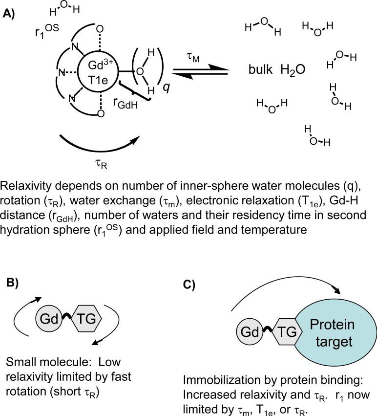
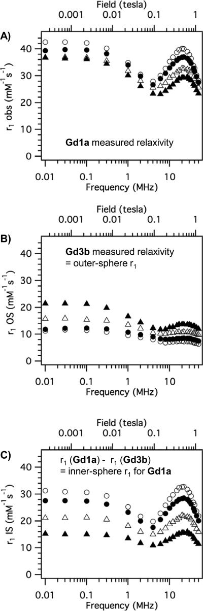
Rationale and objectives: The donor atoms that bind to gadolinium in contrast agents influence inner-sphere water exchange and electronic relaxation, both of which determine observed relaxivity. The effect of these molecular parameters on relaxivity is greatest when the contrast agent is protein bound. We sought to determine an optimal donor atom set to yield high relaxivity compounds.
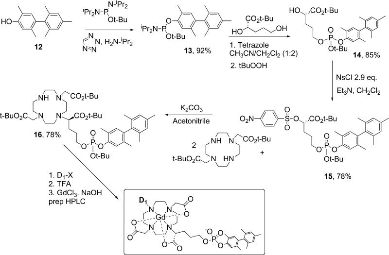
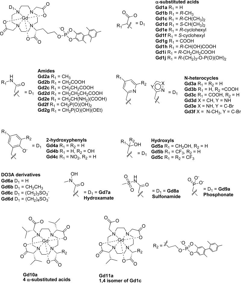
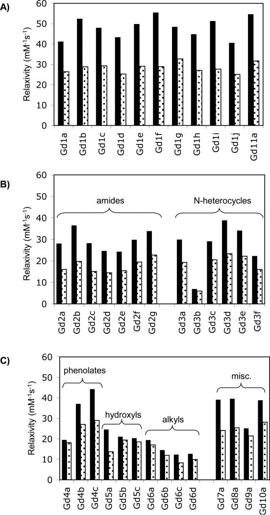
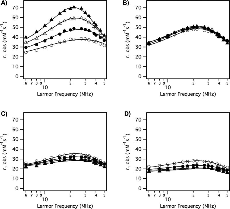
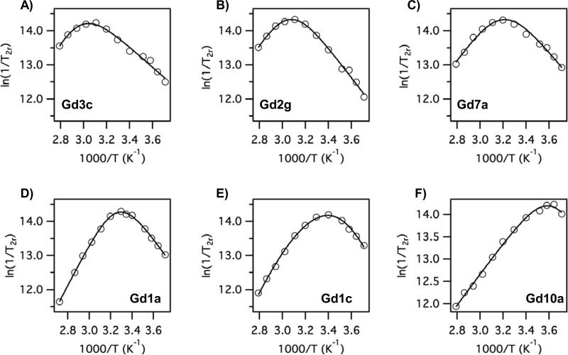
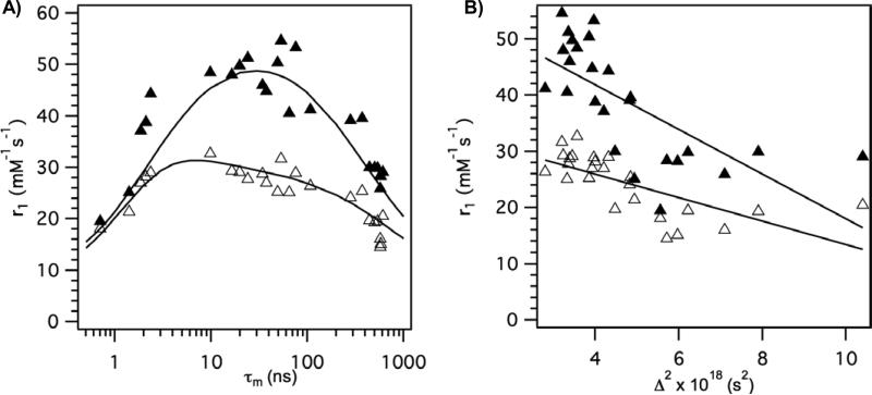
Methods: A total of 38 gadolinium-1,4,7,10-tetraazacyclo-dodecane-N,N',N'',N'''-tetraacetato derivatives were prepared and relaxivity was determined in the presence and absence of human serum albumin as a function of temperature and magnetic field. Each compound had a common albumin-binding group and differed only by substitution of different donor groups at one of the macrocycle nitrogens. Oxygen-17 isotope relaxometry at 7.05 T was performed to estimate water exchange rates.
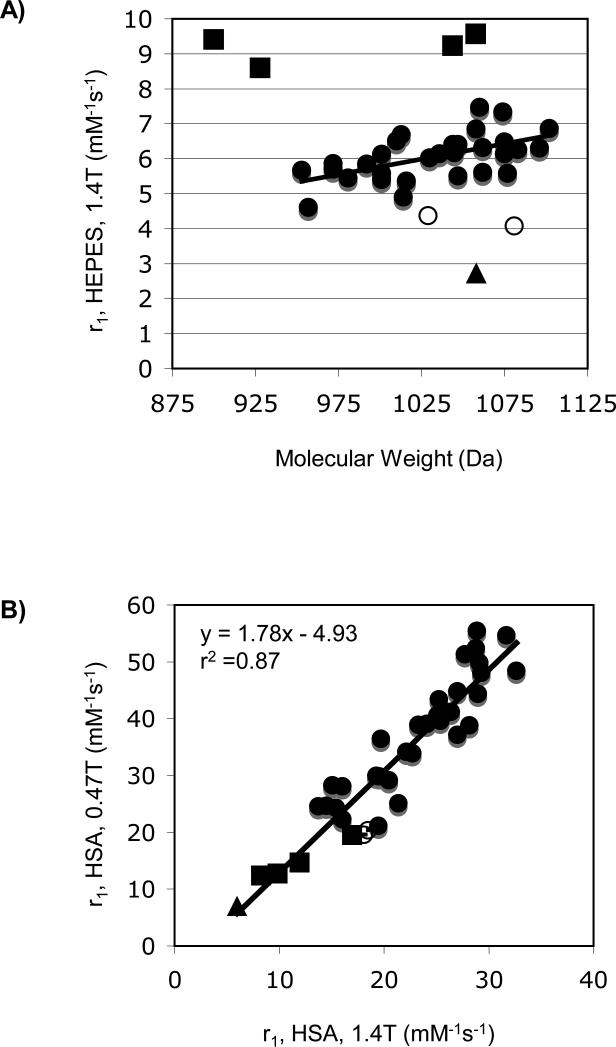
Results: Changing a single donor atom resulted in changes in water exchange rates ranging across 3 orders of magnitude. Donor groups increased water exchange rate in the order: phosphonate ∼ phenolate > α-substituted acetate > acetate > hydroxamate ∼ sulfonamide > amide ∼ pyridyl ∼ imidazole. Relaxivites at 0.47 and 1.4 T, 37°C, ranged from 12.3 to 55.6 mM(-1)s(-1) and from 8.3 to 32.6 mM(-1)s(-1) respectively. Optimal relaxivities were observed when the donor group was an α-substituted acetate. Electronic relaxation was slowest for the acetate derivatives as well.
Conclusions: Water exchange dynamics and relaxivity can be predictably tuned by choice of donor atoms.
Figures
References
-
- Grobner T, Prischl FC. Gadolinium and nephrogenic systemic fibrosis. Kidney Int. 2007;72:260–264. - PubMed
-
- Rydahl C, Thomsen HS, Marckmann P. High Prevalence of Nephrogenic Systemic Fibrosis in Chronic Renal Failure Patients Exposed to Gadodiamide, a Gadolinium-Containing Magnetic Resonance Contrast Agent. Invest Radiol. 2008;43:141–144. - PubMed
-
- Haylor J, Dencausse A, Vickers M, et al. Nephrogenic Gadolinium Biodistribution and Skin Cellularity Following a Single Injection of Omniscan in the Rat. Invest Radiol. 2010;45:XXX–XXX. - PubMed
-
- Aime S, Castelli DD, Crich SG, et al. Pushing the sensitivity envelope of lanthanide-based magnetic resonance imaging (MRI) contrast agents for molecular imaging applications. Acc Chem Res. 2009;42:822–831. - PubMed
Publication types
MeSH terms
Substances
Grants and funding
LinkOut - more resources
Full Text Sources
Other Literature Sources
Medical
