

Thyroid hormone may regulate mRNA abundance in liver by acting on microRNAs
- PMID: 20808432
- PMCID: PMC2921333
- DOI: 10.1371/journal.pone.0012136
Thyroid hormone may regulate mRNA abundance in liver by acting on microRNAs
Abstract
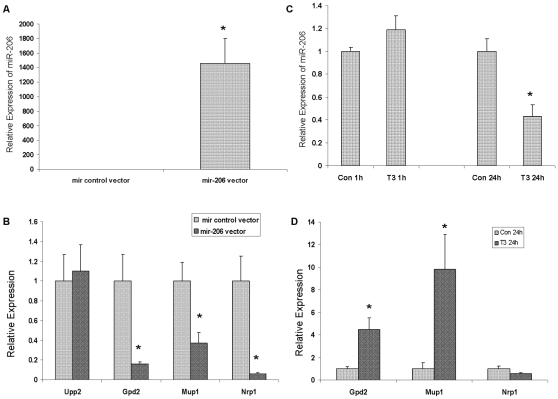
MicroRNAs (miRNAs) are extensively involved in diverse biological processes. However, very little is known about the role of miRNAs in mediating the action of thyroid hormones (TH). Appropriate TH levels are known to be critically important for development, differentiation and maintenance of metabolic balance in mammals. We induced transient hypothyroidism in juvenile mice by short-term exposure to methimazole and perchlorate from post natal day (PND) 12 to 15. The expression of miRNAs in the liver was analyzed using Taqman Low Density Arrays (containing up to 600 rodent miRNAs). We found the expression of 40 miRNAs was significantly altered in the livers of hypothyroid mice compared to euthyroid controls. Among the miRNAs, miRs-1, 206, 133a and 133b exhibited a massive increase in expression (50- to 500-fold). The regulation of TH on the expression of miRs-1, 206, 133a and 133b was confirmed in various mouse models including: chronic hypothyroid, short-term hyperthyroid and short-term hypothyroid followed by TH supplementation. TH regulation of these miRNAs was also confirmed in mouse hepatocyte AML 12 cells. The expression of precursors of miRs-1, 206, 133a and 133b were examined in AML 12 cells and shown to decrease after TH treatment, only pre-mir-206 and pre-mir-133b reached statistical significance. To identify the targets of these miRNAs, DNA microarrays were used to examine hepatic mRNA levels in the short-term hypothyroid mouse model relative to controls. We found transcripts from 92 known genes were significantly altered in these hypothyroid mice. Web-based target predication software (TargetScan and Microcosm) identified 14 of these transcripts as targets of miRs-1, 206, 133a and 133b. The vast majority of these mRNA targets were significantly down-regulated in hypothyroid mice, corresponding with the up-regulation of miRs-1, 206, 133a and 133b in hypothyroid mouse liver. To further investigate target genes, miR-206 was over-expressed in AML 12 cells. TH treatment of cells over-expressing miR-206 resulted in decreased miR-206 expression, and a significant increase in two predicted target genes, Mup1 and Gpd2. The results suggest that TH regulation of these genes may occur secondarily via miR-206. These studies provide new insight into the role of miRNAs in mediating TH regulation of gene expression.
Conflict of interest statement
Figures


Similar articles
-
Hepatic gene expression changes in hypothyroid juvenile mice: characterization of a novel negative thyroid-responsive element.Endocrinology. 2007 Aug;148(8):3932-40. doi: 10.1210/en.2007-0452. Epub 2007 Apr 26. Endocrinology. 2007. PMID: 17463053
-
Temporal analysis of reciprocal miRNA-mRNA expression patterns predicts regulatory networks during differentiation in human skeletal muscle cells.Physiol Genomics. 2015 Mar;47(3):45-57. doi: 10.1152/physiolgenomics.00037.2014. Epub 2014 Dec 29. Physiol Genomics. 2015. PMID: 25547110 Free PMC article.
-
Thyroid hormone negatively regulates CDX2 and SOAT2 mRNA expression via induction of miRNA-181d in hepatic cells.Biochem Biophys Res Commun. 2013 Nov 1;440(4):635-9. doi: 10.1016/j.bbrc.2013.09.116. Epub 2013 Oct 5. Biochem Biophys Res Commun. 2013. PMID: 24103759
-
microRNA-1/133a and microRNA-206/133b clusters: dysregulation and functional roles in human cancers.Oncotarget. 2012 Jan;3(1):9-21. doi: 10.18632/oncotarget.424. Oncotarget. 2012. PMID: 22308266 Free PMC article. Review.
-
MicroRNAs and thyroid hormone action.Mol Cell Endocrinol. 2021 Apr 5;525:111175. doi: 10.1016/j.mce.2021.111175. Epub 2021 Jan 27. Mol Cell Endocrinol. 2021. PMID: 33515639 Review.
Cited by
-
Noncoding RNA Transcripts during Differentiation of Induced Pluripotent Stem Cells into Hepatocytes.Stem Cells Int. 2018 Aug 19;2018:5692840. doi: 10.1155/2018/5692840. eCollection 2018. Stem Cells Int. 2018. PMID: 30210551 Free PMC article.
-
Levothyroxine enhances glucose clearance and blunts the onset of experimental type 1 diabetes mellitus in mice.Br J Pharmacol. 2017 Nov;174(21):3795-3810. doi: 10.1111/bph.13975. Epub 2017 Sep 20. Br J Pharmacol. 2017. PMID: 28800677 Free PMC article.
-
Cloning and identification of a novel thyroid hormone receptor β isoform expressed in the pituitary gland.Mol Cell Biochem. 2014 Apr;389(1-2):141-50. doi: 10.1007/s11010-013-1935-9. Epub 2014 Jan 31. Mol Cell Biochem. 2014. PMID: 24481752
-
An Emerging Role of micro-RNA in the Effect of the Endocrine Disruptors.Front Neurosci. 2016 Jun 30;10:318. doi: 10.3389/fnins.2016.00318. eCollection 2016. Front Neurosci. 2016. PMID: 27445682 Free PMC article. Review.
-
MicroRNAs from plants to animals, do they define a new messenger for communication?Nutr Metab (Lond). 2018 Oct 1;15:68. doi: 10.1186/s12986-018-0305-8. eCollection 2018. Nutr Metab (Lond). 2018. PMID: 30302122 Free PMC article. Review.
References
-
- Boelaert K, Franklyn JA. Thyroid hormone in health and disease. J Endocrinol. 2005;187:1–15. - PubMed
-
- Dong H, Yauk CL, Williams A, Lee A, Douglas GR, et al. Hepatic gene expression changes in hypothyroid juvenile mice: characterization of a novel negative thyroid-responsive element. Endocrinology. 2007;148:3932–3940. - PubMed
-
- Lynn FC. Meta-regulation: microRNA regulation of glucose and lipid metabolism. Trends Endocrinol Metab. 2009;20:452–459. - PubMed
Publication types
MeSH terms
Substances
Grants and funding
LinkOut - more resources
Full Text Sources
Molecular Biology Databases
