. 2010 Mar;20(2):83-91.
doi: 10.1055/s-0029-1242193.

Transcondylar fossa (supracondylar transjugular tubercle) approach: anatomic basis for the approach, surgical procedures, and surgical experience

Affiliations

Transcondylar fossa (supracondylar transjugular tubercle) approach: anatomic basis for the approach, surgical procedures, and surgical experience

Toshio Matsushima et al. Skull Base. 2010 Mar.

Abstract

The authors clarify the anatomic basis and the usefulness of the transcondylar fossa approach (T-C-F A), in which the posterior portion of the jugular tubercle is removed extradurally through the condylar fossa with the atlanto-occipital joint intact. The authors first performed an anatomic study to identify the area to be removed using cadaveric specimens and then applied the T-C-F A to foramen magnum surgeries. The surgeries included clipping a vertebral artery-posterior inferior cerebellar artery aneurysm in 11 cases, microvascular decompression for glossopharyngeal neuralgia in 15 cases, and removing intradural foramen magnum tumors in 17 cases. Only the condylar fossa was removed, but the approach offered very good visualization of the lateral part of the foramen magnum and sufficient working space. These surgeries were performed safely without major complications. This skull base approach is minimally invasive and is not difficult. Therefore, it can be a standard approach for accessing intradural lesions of the foramen magnum. It can be combined with the transcerebellomedullary fissure approach from the lateral side and can also be easily changed to the transcondylar approach, if necessary.

Keywords: Foramen magnum; VA-PICA aneurysm; foramen magnum tumor; glossopharyngeal neuralgia; lateral approaches.


Figures

Figure 1
Figure 1
The lateral wall of the foramen magnum, left side. (A) The bony structure, posterior view with the skull removed partially. The occipital bone has been partially removed, and the posterior condylar canal, jugular tubercle, hypoglossal canal, and condyle are all clearly visible. The jugular tubercle is situated superior to the hypoglossal canal and the condyle is inferior to it. The posterior condylar canal can be seen on the drilled surface of the posterior portion of the jugular tubercle. Oblique lines demonstrate removal of the bone with the transcondylar fossa approach. The green arrow indicates the transcondylar fossa approach. (B) Skull showing the condylar fossa and the condyle, left posteroinferior view. The skull is tilted a little anteriorly and the lateral portion of the foramen magnum is observed. The left condylar fossa, which is located posterior to the occipital condyle, is clearly visible. The posterior condylar canal can also be seen at the bottom of the fossa. (C) The craniovertebral junction. Posterior view in the cadaveric specimen. The occipital bone has been partially removed, and the condyle and atlanto-occipital joint are clearly visible. The large posterior condylar emissary vein runs in the posterior condylar canal, which is located in the bone just posterior to the jugular tubercle. The sigmoid sinus courses laterally and the marginal sinus runs medially. (D) Lateral wall of the foramen magnum at the level of the hypoglossal canal, superior view in a cross section of the cadaveric specimen. The jugular bulb, hypoglossal sinus, and posterior condylar emissary vein can be seen. The vein courses in the posterior portion of the jugular tubercle. (E) A schematic illustration showing the venous system in the left lateral wall of the foramen magnum, superior view. (F) A schematic illustration indicating the bone removed through the left transcondylar fossa approach, superior view. The oblique lines indicate removal of the posterior portion of the jugular tubercle in the approach. A-O, atlanto-occipital; Cond., condylar; Em., emissary; Inf., inferior; Jug., jugular; Marg., marginal; Pet., Petrosal; Post., posterior; V., vein.
Figure 2
Figure 2
Schematic illustrations showing step-by-step surgical procedures of the transcondylar fossa approach. (A) Skin incision and the burr hole making. (B) Bony opening. (C) Sigmoid-magnum triangle, the operative view just after the bony opening, posterior view. The cutting surface of the posterior condylar emissary vein can be seen at the bottom of the condylar fossa. When the dura mater covering the cerebellum is detached and retracted, an exposed bone looks like triangle between the sigmoid sinus and the foramen magnum. (D) Removing the jugular tubercle. The condylar fossa has been drilled and the posterior portion of the jugular tubercle is removed with the atlanto-occipital joint intact, and then the dura mater of the hypoglossal canal is exposed. (E) Incision of dural opening. (F) Intracranial view in a case of meningioma. A-O, atlanto-occipital; Dent. Lig., dentate ligament; Jug., jugular; P.I.C.A., posterior inferior cerebellar artery; Post. Cond. Em. V., posterior condylar emissary vein; V.A., vetebral artery.
Figure 3
Figure 3
A case of unruptured vertebral artery (VA)–posterior inferior cerebellar artery (PICA) aneurysm, left. (A) Preoperative three-dimensional computed tomography angiography (3D-CTA), posterosuperior view, demonstrating a saccular aneurysm (red arrow) at the left VA-PICA junction. (B) Intraoperative microscopic view, posterior view. The saccular aneurysm can be seen behind the PICA. The VA and the cranial nerve 11th and 12th are clearly visible. (C) Postoperative 3D-CTA, posterior view, demonstrating the aneurismal clip (green arrow) parallel to the VA and the drilled jugular tubercle (red arrow). PICA, posterior inferior cerebellar artery; VA, vertebral artery.
Figure 4
Figure 4
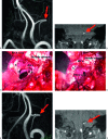
A case of glossopharyngeal neuralgia, left. (A) Preoperative magnetic resonance angiography (MRA), anterior view, demonstrating the loop of the posterior inferior cerebellar artery (PICA; red arrow) with a high origin. (B) Preoperative magnetic resonance imaging (MRI), anterior view, demonstrating the loop of the PICA (red arrow) in the supraolivary fossette. (C) Intraoperative microscopic view, posterior view. The loop of the PICA is situated between the 9th cranial nerve and the 10th cranial nerve. (D) Intraoperative microscopic view. The loop of the PICA has been pulled out. (E) Postoperative MRA, anterior view, demonstrating the shift of the loop of the PICA. (F) Postoperative MRI, anterior view, demonstrating the shift of the PICA in the supraolivary fossette. PICA, posterior inferior cerebellar artery.
Figure 5
Figure 5
A case of recurrent meningioma growing into the fourth ventricle. (A) Preoperative enhanced magnetic resonance imaging (MRI), coronal view, demonstrating the tumor (red arrow). (B) Preoperative enhanced MRI, axial view, demonstrating the tumor (red arrow) growing into the fourth ventricle. (C) Postoperative enhanced MRI, coronal view, demonstrating the cavity after removing the tumor. (D) Postoperative enhanced MRI, axial view, demonstrating subtotal removal of the tumor.

Similar articles

Cited by

  • Quantitative Anatomical Study of Tailored Far-Lateral Approach for the VA-PICA Regions.
    Kim YD, Mendes GA, Seoane P, Agrawal A, Maramreddy N, Nakaji P, Spetzler RF, Preul MC. Kim YD, et al. J Neurol Surg B Skull Base. 2015 Feb;76(1):57-65. doi: 10.1055/s-0034-1389373. Epub 2014 Sep 21. J Neurol Surg B Skull Base. 2015. PMID: 25685651 Free PMC article.
  • Surgery of the lateral skull base: a 50-year endeavour.
    Zanoletti E, Mazzoni A, Martini A, Abbritti RV, Albertini R, Alexandre E, Baro V, Bartolini S, Bernardeschi D, Bivona R, Bonali M, Borghesi I, Borsetto D, Bovo R, Breun M, Calbucci F, Carlson ML, Caruso A, Cayé-Thomasen P, Cazzador D, Champagne PO, Colangeli R, Conte G, D'Avella D, Danesi G, Deantonio L, Denaro L, Di Berardino F, Draghi R, Ebner FH, Favaretto N, Ferri G, Fioravanti A, Froelich S, Giannuzzi A, Girasoli L, Grossardt BR, Guidi M, Hagen R, Hanakita S, Hardy DG, Iglesias VC, Jefferies S, Jia H, Kalamarides M, Kanaan IN, Krengli M, Landi A, Lauda L, Lepera D, Lieber S, Lloyd SLK, Lovato A, Maccarrone F, Macfarlane R, Magnan J, Magnoni L, Marchioni D, Marinelli JP, Marioni G, Mastronardi V, Matthies C, Moffat DA, Munari S, Nardone M, Pareschi R, Pavone C, Piccirillo E, Piras G, Presutti L, Restivo G, Reznitsky M, Roca E, Russo A, Sanna M, Sartori L, Scheich M, Shehata-Dieler W, Soloperto D, Sorrentino F, Sterkers O, Taibah A, Tatagiba M, Tealdo G, Vlad D, Wu H, Zanetti D. Zanoletti E, et al. Acta Otorhinolaryngol Ital. 2019 Jun;39(SUPPL. 1):S1-S146. doi: 10.14639/0392-100X-suppl.1-39-2019. Acta Otorhinolaryngol Ital. 2019. PMID: 31130732 Free PMC article. Review.
  • Course of the V3 segment of the vertebral artery relative to the suboccipital triangle as an anatomical marker for a safe far lateral approach: A retrospective clinical study.
    Sriamornrattanakul K, Akharathammachote N, Chonhenchob A, Mongkolratnan A, Niljianskul N, Phoominaonin IS, Ariyaprakai C, Wongsuriyanan S. Sriamornrattanakul K, et al. Surg Neurol Int. 2022 Jul 15;13:304. doi: 10.25259/SNI_346_2022. eCollection 2022. Surg Neurol Int. 2022. PMID: 35928311 Free PMC article.
  • Midline suboccipital approach to a vertebral artery-posterior inferior cerebellar artery aneurysm from the rostral end of the patient using ORBEYE.
    Murakami T, Toyota S, Nakagawa K, Hagioka T, Hoshikuma Y, Suematsu T, Shimizu T, Kobayashi M, Taki T. Murakami T, et al. Surg Neurol Int. 2022 Mar 11;13:87. doi: 10.25259/SNI_1272_2021. eCollection 2022. Surg Neurol Int. 2022. PMID: 35399900 Free PMC article.
  • Multimodal management of giant solid hemangioblastomas in two patients with preoperative embolization.
    Vargas-Urbina J, Crisanto-Silva JA, Vásquez-Perez C, Davila-Adrianzén A, Alcas-Seminario D, Lines-Aguilar W, Mamani-Choquepata R, Panta-Rojas G. Vargas-Urbina J, et al. Surg Neurol Int. 2024 Apr 26;15:144. doi: 10.25259/SNI_28_2024. eCollection 2024. Surg Neurol Int. 2024. PMID: 38742001 Free PMC article.

References

    1. Bertalanffy H, Seeger W. The dorsolateral, suboccipital, transcondylar approach to the lower clivus and anterior portion of the craniocervical junction. Neurosurgery. 1991;29:815–821. - PubMed
    1. Gilsbach J M, Sure U, Mann W. The supracondylar approach to the jugular tubercle and hypoglossal canal. Surg Neurol. 1998;50:563–570. - PubMed
    1. Hakuba A. In: Sekhar LN, Janecka IP, editor. Surgery of Cranial Base Tumors. New York: Raven Press; 1993. Tsujimoto: Transcondyle approach for foramen magnum meningiomas. pp. 671–678.
    1. Heros R C. Lateral suboccipital approach for vertebral and vertebrobasilar artery lesions. J Neurosurg. 1986;64:559–562. - PubMed
    1. Margalit N S, Lesser J B, Singer M, Sen C. Lateral approach to anterolateral tumors at the foramen magnam: factors determining surgical procedure. Neurosurgery. 2005;56(2 Suppl):324–336. - PubMed