. 2010 Mar;8(1):26-40.
doi: 10.2174/157015910790909511.

Neuropharmacology of vestibular system disorders

Affiliations

Neuropharmacology of vestibular system disorders

Enrique Soto et al. Curr Neuropharmacol. 2010 Mar.

Abstract

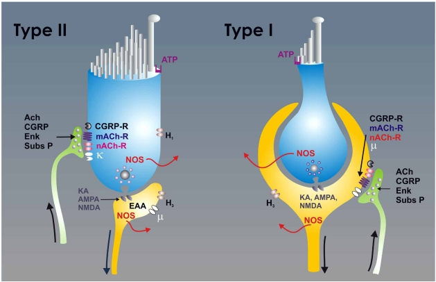
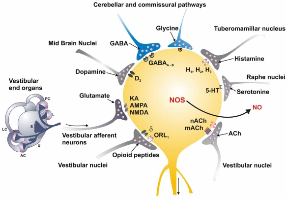
This work reviews the neuropharmacology of the vestibular system, with an emphasis on the mechanism of action of drugs used in the treatment of vestibular disorders. Otolaryngologists are confronted with a rapidly changing field in which advances in the knowledge of ionic channel function and synaptic transmission mechanisms have led to the development of new scientific models for the understanding of vestibular dysfunction and its management. In particular, there have been recent advances in our knowledge of the fundamental mechanisms of vestibular system function and drug mechanisms of action. In this work, drugs acting on vestibular system have been grouped into two main categories according to their primary mechanisms of action: those with effects on neurotransmitters and neuromodulator receptors and those that act on voltage-gated ion channels. Particular attention is given in this review to drugs that may provide additional insight into the pathophysiology of vestibular diseases. A critical review of the pharmacology and highlights of the major advances are discussed in each case.

Keywords: Dizziness; Inner ear; Ménière's disease; Vertigo; excitatory amino acids.; hair cells; vestibular nuclei.


Figures

Fig. (1)
Fig. (1)
The scheme depicts the synaptic relationships of type I (right) and type II (left) hair cells. The type I hair cells are characterized by the large chaliceal afferent innervation that covers its basolateral surface. The efferent fibers make synaptic contacts with the external surface of the calyx in the afferent neuron. In type II hair cells, the afferent neurons form button type synapses, and the efferent neurons make synaptic contact directly upon the hair cell body. The hair cell to afferent synapse uses glutamate as the principal neurotransmitter. At the postsynaptic cell glutamate interacts with several subtypes of excitatory amino acid (EAA)-receptors including N-methyl-D-aspartic acid (NMDA), α-amino-3-hydroxyl-5-methyl-4-isoxazole-propionic acid (AMPA), kainic acid (KA) and metabotropic receptors. The efferent neurons are primarily cholinergic, and ACh released from afferent neurons interacts with muscarinic (mACh) and nicotinic (nACh) receptors. The efferent neurons also release calcitonin gene related peptide (CGRP), substance-P and enkephalins, which act on specific receptors (in the case of the enkephalins κ−opioid receptor in the hair cells and µ−opioid receptors in the afferent neurons). Both the hair cells and the afferent neurons expressed the nitric oxide synthase (NOS) and produced nitric oxide (NO). Hair cells also express H1 histamine receptors and the afferent neurons H3 histamine receptors. The hair cells typically express purinergic receptors (ATP) in their apical portion.
Fig. (2)
Fig. (2)
The scheme depicts the complexity of synaptic input impinging on the vestibular nucleus neurons. It is necessary to consider that the neurons of the nuclei are heterogeneous and not all cells necessarily receive all types of synaptic influences. The main synaptic input to the vestibular nuclei neurons is from the primary afferents, mediated by glutamate that interacts with NMDA, AMPA/KA and metabotropic receptors. Vestibular nuclei also receive glutamatergic synapses originating from spinal cord neurons. GABAergic fibers originating primarily from the cerebellum and from the contralateral vestibular nuclei impinge on the vestibular nucleus neurons, activating GABA-A and GABA-B receptors. Histaminergic fibers originating from the tuberomammillar nucleus act on H1, H2 and H3 receptors. Serotonergic fibers originating from the raphe nuclei activate 5-HT1 and 5-HT2 receptors. Intrinsic and commissural connections give rise to glycinergic fibers acting on glycine inhibitory receptors. Noradrenergic fibers originating from the locus coeruleus act primarily on α2 receptors, but also on α1 and β receptors. Internuclear enkephalinergic fibers that release orphanin-nociceptin F/Q acting on ORL1 receptors (opioid-like orphan receptor) and δ-opioid receptors also make a synaptic input to certain neurons in the nuclei. The endocannabinergic type CB1 receptors have also been detected in the vestibular nuclei. The output of the vestibular nuclei neurons is primarily by glutamatergic and cholinergic projections, but GABAergic and glycinergic projections have been demonstrated also. Finally, the vestibular nucleus neurons express the NOS and may produce NO as a cellular messenger.

Similar articles

Cited by

References

    1. Aantaa E. Treatment of acute vestibular vertigo. Acta Otolaryngol. Suppl. 1991;479:44–47. - PubMed
    1. Adunka O, Moustaklis E, Weber A, May A, von Ilberg C, Gstoettner W, Kierner AC. Labyrinth anesthesia-a forgotten but practical treatment option in Ménière's disease. ORL J. Otorhinolaryngol. Relat. Spec. 2003;65:84–90. - PubMed
    1. Alacio Casero J, Ortega del Álamo P. Efectividad de trimetazidina en pacientes con alteraciones del equilibrio. Estudio prospectivo multicéntrico. ORL-DIPS. 2003;30:184–192.
    1. Almanza A, Vega R, Soto E. Calcium current in type I hair cells isolated from the semicircular canal crista ampullaris of the rat. Brain Res. 2003;994:175–180. - PubMed
    1. Anderson AD, Troyanovskaya M, Wackym PA. Differential expression of alpha2-7, alpha9 and beta2-4 nicotinic acetylcholine receptor subunit mRNA in the vestibular end-organs and Scarpa's ganglia of the rat. Brain Res. 1997;778:409–413. - PubMed

LinkOut - more resources