

Femoroacetabular impingement syndrome: a narrative review for the chiropractor
- PMID: 20808616
- PMCID: PMC2921782
Femoroacetabular impingement syndrome: a narrative review for the chiropractor
Abstract
Objective: To familiarize the chiropractic clinician with the clinical presentation, radiographic features, and conservative versus surgical treatment options for managing femoroacetabular impingement (FAI) syndrome.
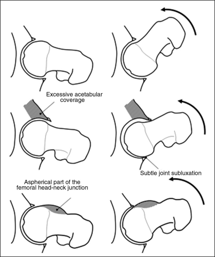
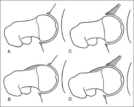
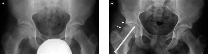
Background: FAI syndrome is a relatively new clinical entity to be described in orthopedics, and has been strongly linked with pain and early osteoarthritis of the hip in young adults. Hip joint radiographs in these patients often appear normal at first-particularly if the clinician is unfamiliar with FAI. The role of conservative therapy in managing this disorder is questionable. Surgical treatment ultimately addresses any acetabular labral or articular cartilage damage, as well as the underlying osseous abnormalities associated with FAI. The most commonly used approach is open surgical hip dislocation; however, more recent surgical procedures also involve arthroscopy.
Conclusion: In FAI syndrome-a condition unknown to many clinicians (including medical)-chiropractors can play an important role in its diagnosis and referral for appropriate management.
Objectif :: Familiariser le chiropraticien clinicien avec la présentation clinique, les caractéristiques radiographiques, et les options de traitement conservateur par opposition aux traitements chirurgicaux dans la gestion du syndrome du conflit fémoroacétabulaire.
Contexte :: Le syndrome du conflit fémoroacétabulaire est une entité clinique dont la description orthopédique est relativement récente et qui a été fortement mise en lien avec la douleur et l’arthrose précoce de la hanche chez les jeunes adultes. Des radiographies de l’articulation de la hanche de ces patients apparaissent souvent normales a priori, surtout lorsque le clinicien n’est pas familiarisé avec le syndrome du conflit fémoroacétabulaire. Le rôle d’une thérapie conservatrice dans la gestion de ce trouble est discutable. Le traitement chirurgical aborde ultimement tout dommage du cotyle labial ou du cartilage de l’articulation, en plus des anormalités osseuses sous-jacentes associées au syndrome du conflit fémoroacétabulaire. L’approche la plus communément employée est la luxation chirurgicale effractive de la hanche. Toutefois, des procédures chirurgicales récentes emploient également l’arthroscopie.
Conclusion :: En ce qui concerne le syndrome du conflit fémoroacétabulaire, un trouble inconnu de plusieurs cliniciens (y compris le personnel médical), les chiropraticiens peuvent jouer un rôle important sur le plan du diagnostic et du renvoi vers une gestion convenable.
Keywords: acetabulum/abnormalities; femoral neck/abnormalities; hip joint; osteoarthritis.
Figures
Similar articles
-
What Are the Risk Factors for Revision Surgery After Hip Arthroscopy for Femoroacetabular Impingement at 7-year Followup?Clin Orthop Relat Res. 2017 Apr;475(4):1169-1177. doi: 10.1007/s11999-016-5115-6. Clin Orthop Relat Res. 2017. PMID: 27718121 Free PMC article.
-
The Pattern of Acetabular Cartilage Wear Is Hip Morphology-dependent and Patient Demographic-dependent.Clin Orthop Relat Res. 2019 May;477(5):1021-1033. doi: 10.1097/CORR.0000000000000649. Clin Orthop Relat Res. 2019. PMID: 30998630 Free PMC article.
-
Labral Reattachment in Femoroacetabular Impingement Surgery Results in Increased 10-year Survivorship Compared With Resection.Clin Orthop Relat Res. 2017 Apr;475(4):1178-1188. doi: 10.1007/s11999-016-5114-7. Clin Orthop Relat Res. 2017. PMID: 27744594 Free PMC article.
-
Femoroacetabular impingement: an arthroscopic solution.Bone Joint J. 2013 Nov;95-B(11 Suppl A):26-30. doi: 10.1302/0301-620X.95B11.33016. Bone Joint J. 2013. PMID: 24187347 Review.
-
MRI for the preoperative evaluation of femoroacetabular impingement.Insights Imaging. 2016 Apr;7(2):187-98. doi: 10.1007/s13244-015-0459-0. Epub 2015 Dec 29. Insights Imaging. 2016. PMID: 26715128 Free PMC article. Review.
Cited by
-
NON-OPERATIVE MANAGEMENT OF INDIVIDUALS WITH NON-ARTHRITIC HIP PAIN: A LITERATURE REVIEW.Int J Sports Phys Ther. 2019 Feb;14(1):135-147. Int J Sports Phys Ther. 2019. PMID: 30746300 Free PMC article.
-
Femoroacetabular Impingement: A Retrospective Case Study With 8-Year Follow-Up.J Chiropr Med. 2015 Dec;14(4):290-6. doi: 10.1016/j.jcm.2015.05.006. Epub 2015 Nov 18. J Chiropr Med. 2015. PMID: 26793042 Free PMC article.
-
Femoroacetabular impingement: is hyaluronic acid effective?Knee Surg Sports Traumatol Arthrosc. 2014 Apr;22(4):889-92. doi: 10.1007/s00167-013-2581-1. Epub 2013 Jun 28. Knee Surg Sports Traumatol Arthrosc. 2014. PMID: 23812440 Clinical Trial.
-
Advanced hip joint degeneration associated with femoroacetabular impingement in a retired chiropractor.J Can Chiropr Assoc. 2016 Sep;60(3):260-262. J Can Chiropr Assoc. 2016. PMID: 27713583 Free PMC article.
-
Can the Open Stance Forehand Increase the Risk of Hip Injuries in Tennis Players?Orthop J Sports Med. 2020 Dec 11;8(12):2325967120966297. doi: 10.1177/2325967120966297. eCollection 2020 Dec. Orthop J Sports Med. 2020. PMID: 33354579 Free PMC article.
References
-
- Ganz R, Parvizi J, Beck M, Leunig M, Nötzli H, Siebenrock KA. Femoroacetabular impingement: a cause for osteoarthritis of the hip. Clin Orthop Relat Res. 2003;417:112–120. - PubMed
-
- Beck M, Kalhor M, Leunig M, Ganz R. Hip morphology influences the pattern of damage to the acetabular cartilage: femoroacetabular impingement as a cause of early osteoarthritis of the hip. J Bone Joint Surg Br. 2005;87(7):1012–1018. - PubMed
LinkOut - more resources
Full Text Sources