

Laser capture microdissection in the tissue biorepository
- PMID: 20808641
- PMCID: PMC2922833
Laser capture microdissection in the tissue biorepository
Abstract
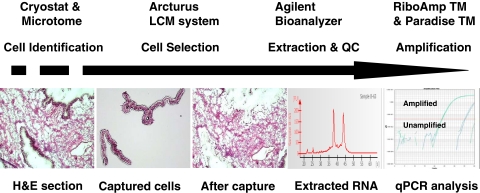
An important need of many cancer research projects is the availability of high-quality, appropriately selected tissue. Tissue biorepositories are organized to collect, process, store, and distribute samples of tumor and normal tissue for further use in fundamental and translational cancer research. This, in turn, provides investigators with an invaluable resource of appropriately examined and characterized tissue specimens and linked patient information. Human tissues, in particular, tumor tissues, are complex structures composed of heterogeneous mixtures of morphologically and functionally distinct cell types. It is essential to analyze specific cell types to identify and define accurately the biologically important processes in pathologic lesions. Laser capture microdissection (LCM) is state-of-the-art technology that provides the scientific community with a rapid and reliable method to isolate a homogeneous population of cells from heterogeneous tissue specimens, thus providing investigators with the ability to analyze DNA, RNA, and protein accurately from pure populations of cells. This is particularly well-suited for tumor cell isolation, which can be captured from complex tissue samples. The combination of LCM and a tissue biorepository offers a comprehensive means by which researchers can use valuable human biospecimens and cutting-edge technology to facilitate basic, translational, and clinical research. This review provides an overview of LCM technology with an emphasis on the applications of LCM in the setting of a tissue biorepository, based on the author's extensive experience in LCM procedures acquired at Fox Chase Cancer Center and Hollings Cancer Center.
Keywords: cancer biology; cells of interest; pathology.
Figures
References
-
- Monajembashi S, Cremer C, Cremer T, Wolfrum J, Greulich KO. Microdissection of human chromosomes by a laser microbeam. Exp Cell Res 1986;167:262–265 - PubMed
-
- Emmert-Buck MR, Bonner RF, Smith PD, et al. Laser capture microdissection. Science 1996;274:998–1001 - PubMed
-
- Anoruo B, van Oorschot R, Mitchell J, Howells D. Isolating cells from non-sperm cellular mixtures using the PALM microlaser micro dissection system. Forensic Sci Int 2007;173:93–96 - PubMed
Publication types
MeSH terms
Substances
LinkOut - more resources
Full Text Sources