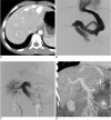
Congenital intrahepatic portosystemic venous shunt and liver mass in a child patient: successful endovascular treatment with an amplatzer vascular plug (AVP)
- PMID: 20808706
- PMCID: PMC2930171
- DOI: 10.3348/kjr.2010.11.5.583
Congenital intrahepatic portosystemic venous shunt and liver mass in a child patient: successful endovascular treatment with an amplatzer vascular plug (AVP)
Abstract
A congenital intrahepatic portosystemic shunt is a rare anomaly; but, the number of diagnosed cases has increased with advanced imaging tools. Symptomatic portosystemic shunts, especially those that include hyperammonemia, should be treated; and various endovascular treatment methods other than surgery have been reported. Hepatic masses with either an intra- or extrahepatic shunt also have been reported, and the mass is another reason for treatment. Authors report a case of a congenital intrahepatic portosystemic shunt with a hepatic mass that was successfully treated using a percutaneous endovascular approach with vascular plugs. By the time the first short-term follow-up was conducted, the hepatic mass had disappeared.
Keywords: Liver neoplasm; Portosystemic shunt, surgical; Radiology, interventional.
Figures

Comment in
-
RE: endovascular treatment of congenital intrahepatic portosystemic shunts with Amplatzer plugs.Korean J Radiol. 2012 Jan-Feb;13(1):115. doi: 10.3348/kjr.2012.13.1.115. Epub 2011 Dec 23. Korean J Radiol. 2012. PMID: 22247647 Free PMC article. No abstract available.
References
-
- Chagnon SF, Vallee CA, Barge J, Chevalier LJ, Le Gal J, Blery MV. Aneurysmal portahepatic venous fistula: report of two cases. Radiology. 1986;159:693–695. - PubMed
-
- Singh K, Kapoor A, Kapoor A, Gupta K, Mahajan G. Congenital intrahepatic portosystemic shunt--an incidental rare anomaly. Indian J Pediatr. 2006;73:1122–1123. - PubMed
-
- Stringer MD. The clinical anatomy of congenital portosystemic venous shunts. Clin Anat. 2008;21:147–157. - PubMed
-
- Kanamori Y, Hashizume K, Kitano Y, Sugiyama M, Motoi T, Tange T. Congenital extrahepatic portocaval shunt (Abernethy type 2), huge liver mass, and patent ductus arteriosus--a case report of its rare clinical presentation in a young girl. J Pediatr Surg. 2003;38:E15. - PubMed
-
- Ikeda S, Sera Y, Yoshida M, Izaki T, Uchino S, Endo F, et al. Successful coil embolization in an infant with congenital intrahepatic portosystemic shunts. J Pediatr Surg. 1999;34:1031–1032. - PubMed
Publication types
MeSH terms
LinkOut - more resources
Full Text Sources
Medical
Miscellaneous

