

doi: 10.1088/1367-2630/12/7/073011.
Design of an electron microscope phase plate using a focused continuous-wave laser
Affiliations
- PMID: 20808709
- PMCID: PMC2929835
- DOI: 10.1088/1367-2630/12/7/073011
Item in Clipboard
Design of an electron microscope phase plate using a focused continuous-wave laser
New J Phys.
2010 Jul.
Abstract
We propose a Zernike phase contrast electron microscope that uses an intense laser focus to convert a phase image into a visible image. We present the relativistic quantum theory of the phase shift caused by the laser-electron interaction, study resonant cavities for enhancing the laser intensity and discuss applications in biology, soft-materials science and atomic and molecular physics.
Figures
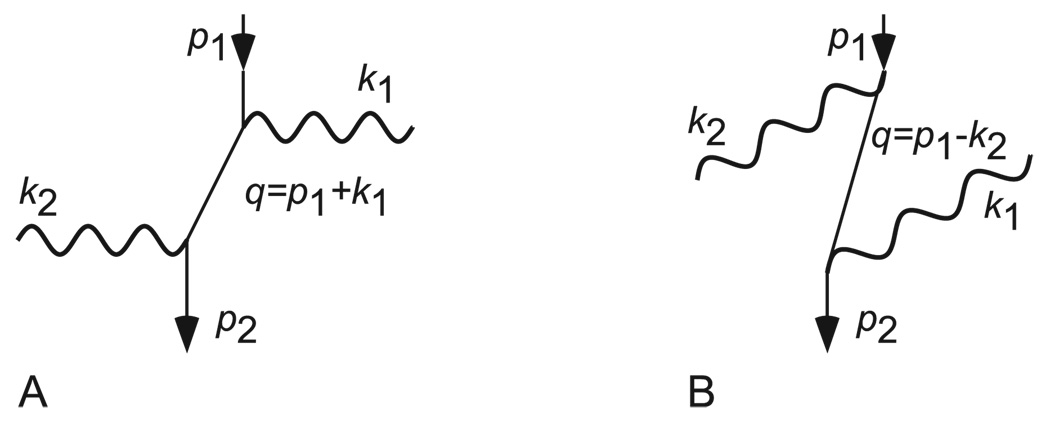
Feynman diagrams for Compton scattering. p1, q, and p2, respectively, denote the initial, intermediate and final momenta of the electron; k1 and k2 are the photon momenta.
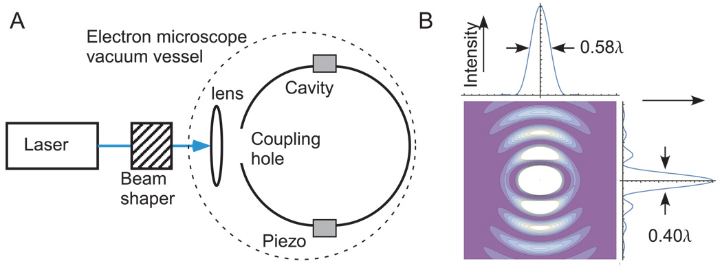
(A) The setup for a phase contrast TEM using a near-spherical resonant cavity. The electron beam is propagating orthogonal to the paper plane. (B) Intensity of the TMn01 mode in a spherical cavity. Shown is a two-dimensional contour plot as well as the intensities along the x and y axes through the focus, respectively.
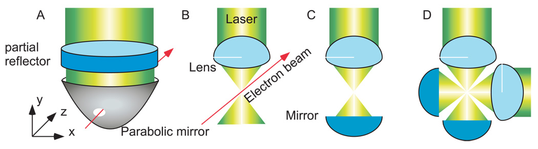
Alternative realizations of laser phase plates. (A) Plano-parabolic cavity; (B) high-NA lens; (C) high-NA lens with retroreflector; (D) two focused, retroreflected beams. The electron beam is propagating orthogonally to the page.
Similar articles
-
Near-concentric Fabry-Pérot cavity for continuous-wave laser control of electron waves.Opt Express. 2017 Jun 26;25(13):14453-14462. doi: 10.1364/OE.25.014453. Opt Express. 2017. PMID: 28789031
-
Observation of the Relativistic Reversal of the Ponderomotive Potential.Phys Rev Lett. 2020 May 1;124(17):174801. doi: 10.1103/PhysRevLett.124.174801. Phys Rev Lett. 2020. PMID: 32412292 Free PMC article.
-
Improved Zernike-type phase contrast for transmission electron microscopy.J Microsc. 2015 Jul;259(1):74-8. doi: 10.1111/jmi.12250. Epub 2015 Apr 9. J Microsc. 2015. PMID: 25865092
-
Strong-laser-field physics, non-classical light states and quantum information science.Rep Prog Phys. 2023 Aug 7;86(9). doi: 10.1088/1361-6633/acea31. Rep Prog Phys. 2023. PMID: 37489874 Review.
-
Phase recovery and lensless imaging by iterative methods in optical, X-ray and electron diffraction.Philos Trans A Math Phys Eng Sci. 2002 May 15;360(1794):875-95. doi: 10.1098/rsta.2001.0972. Philos Trans A Math Phys Eng Sci. 2002. PMID: 12804284 Review.
Cited by
-
High-power near-concentric Fabry-Perot cavity for phase contrast electron microscopy.Rev Sci Instrum. 2021 May 1;92(5):053005. doi: 10.1063/5.0045496. Rev Sci Instrum. 2021. PMID: 34243315 Free PMC article.
-
Optical Excitations with Electron Beams: Challenges and Opportunities.ACS Photonics. 2021 Apr 21;8(4):945-974. doi: 10.1021/acsphotonics.0c01950. Epub 2021 Mar 25. ACS Photonics. 2021. PMID: 35356759 Free PMC article. Review.
-
Could Egg White Lysozyme be Solved by Single Particle Cryo-EM?J Chem Inf Model. 2020 May 26;60(5):2605-2613. doi: 10.1021/acs.jcim.9b01176. Epub 2020 May 11. J Chem Inf Model. 2020. PMID: 32202786 Free PMC article.
-
Volta potential phase plate for in-focus phase contrast transmission electron microscopy.Proc Natl Acad Sci U S A. 2014 Nov 4;111(44):15635-40. doi: 10.1073/pnas.1418377111. Epub 2014 Oct 20. Proc Natl Acad Sci U S A. 2014. PMID: 25331897 Free PMC article.
-
Overcoming resolution loss due to thermal magnetic field fluctuations from phase plates in transmission electron microscopy.Ultramicroscopy. 2023 Jul;249:113730. doi: 10.1016/j.ultramic.2023.113730. Epub 2023 Mar 29. Ultramicroscopy. 2023. PMID: 37011498 Free PMC article.
References
-
- Glaeser R, Downing K, DeRosier D, Chiu W, Frank J. Electron Crystallography of Biological Macromolecules. Oxford: Oxford University Press; 2007.
- Spence JCH. High Resolution Electron Microscopy. New York: Oxford University Press; 2003.
-
- Zernicke F. Physica. 1942;9:686–698.
- Zernicke F. Physica. 1942;9:974–986.
- Zernicke F. Science. 1955;121:345–349. - PubMed
-
- Boersch H. Z. Nat.forsch. A. 1947;2:615.
-
- Hosokawa F, Danev R, Arai Y, Nagayama K. J. Electron Microsc. 2005;54:317–324. - PubMed
Grants and funding
LinkOut - more resources
Full Text Sources
Other Literature Sources