. 2010 Aug 24;5(8):e12353.
doi: 10.1371/journal.pone.0012353.

Colorimetric measurement of triglycerides cannot provide an accurate measure of stored fat content in Drosophila

Affiliations

Colorimetric measurement of triglycerides cannot provide an accurate measure of stored fat content in Drosophila

Bader Al-Anzi et al. PLoS One. .

Abstract

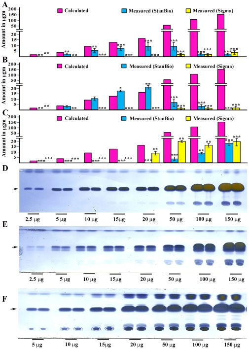
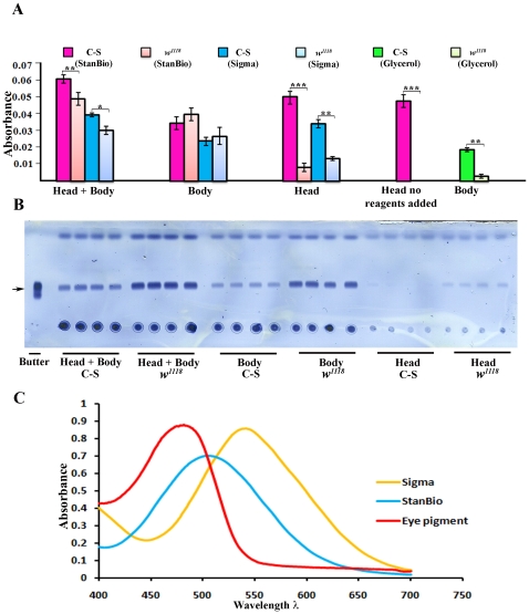
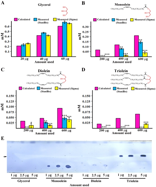
Drosophila melanogaster has recently emerged as a useful model system in which to study the genetic basis of regulation of fat storage. One of the most frequently used methods for evaluating the levels of stored fat (triglycerides) in flies is a coupled colorimetric assay available as a kit from several manufacturers. This is an aqueous-based enzymatic assay that is normally used for measurement of mammalian serum triglycerides, which are present in soluble lipoprotein complexes. In this short communication, we show that coupled colorimetric assay kits cannot accurately measure stored triglycerides in Drosophila. First, they fail to give accurate readings when tested on insoluble triglyceride mixtures with compositions like that of stored fat, or on fat extracted from flies with organic solvents. This is probably due to an inability of the lipase used in the kits to efficiently cleave off the glycerol head group from fat molecules in insoluble samples. Second, the measured final products of the kits are quinoneimines, which absorb visible light in the same wavelength range as Drosophila eye pigments. Thus, when extracts from crushed flies are assayed, much of the measured signal is actually due to eye pigments. Finally, the lipoprotein lipases used in colorimetric assays also cleave non-fat glycerides. The glycerol backbones liberated from all classes of glycerides are measured through the remaining reactions in the assay. As a consequence, when these assay kits are used to evaluate tissue extracts, the observed signal actually represents the amount of free glycerols together with all types of glycerides. For these reasons, findings obtained through use of coupled colorimetric assays on Drosophila samples must be interpreted with caution. We also show here that using thin-layer chromatography to measure stored triglycerides in flies eliminates all of these problems.


Conflict of interest statement

Competing Interests: The authors have declared that no competing interests exist.

Figures

Figure 1
Figure 1. The coupled colorimetric assay is unsuitable for analysis of insoluble triglyceride mixtures.
Lard (A), butter (B), and extracted fly fat (C) do not produce linear increases in quinoneimine absorption as more of each substance is assayed. Both the Stanbio Triglyceride Liquicolor Kit (blue bars) and the Sigma TR0100 kit (yellow bars) were used. The pink bars (Calculated) indicate the signal that would be observed if the samples could be measured accurately. Error bars are standard deviations of four different replicates for a given sample type. The butterfat and lard samples were sonicated and boiled as in ref. . The extracted fly fat samples were resuspended without sonication and boiling, because sonicated and boiled samples generated even smaller signals (data not shown). Lard (D), butter (E), and extracted fly fat (F) all produce signals that increase with increasing amounts of either substance when assayed by the TLC method. The arrows indicate the location of the triglyceride band on the TLC assay. Asterisks denote T-test statistical significance: *; P<0.05–0,005, **; P<0.005–0.0005, ***; P<0.0005.
Figure 2
Figure 2. The coupled colorimetric triglyceride assay cannot accurately measure stored fat in adult Drosophila.
Whole w1118 (VDRC) flies contain more triglyceride than Canton S (C-S) flies, as shown by the stronger triglyceride bands (arrow) in (B) (head+body). Four replicates are shown. However, w1118 produces weaker signals in the colorimetric assay than C-S (A, head+body bars, when either the StanBio or Sigma kits are used). The bars represent an average of the values from four replicates. The TLC assay shows that, as expected, heads have much less fat than bodies. w1118 heads contain more fat than C-S heads (B, head). However, the colorimetric signal from C-S heads is much greater than that from w1118 heads. The same signal strength is observed when C-S heads are assayed without any added enzymes, showing that this signal is due to absorption by eye pigment. w1118 bodies produce a slightly stronger signal than C-S bodies, consistent with the TLC results; but the difference between C-S and w1118 bodies in the colorimetric assay is not statistically significant. We also measured glycerols in C-S and w1118 bodies, using the 'no lipase' reaction mix in the Sigma TR0100 kit. C-S bodies (bright green bars) contain much more glycerol than w1118 bodies (light green bars). Error bars are standard deviations of four different replicates for a given sample type. Asterisks denote T-test statistical significance: *; P<0.05–0,005, **; P<0.005–0.0005, ***; P<0.0005. (C) Absorption spectra of quinoneimines produced by the StanBio and Sigma kits, and of fly eye pigment.
Figure 3
Figure 3. Coupled colorimetric assays cannot distinguish triglyceride from other glycerides.
Glycerol (a soluble compound) can be accurately measured using either kit (A). Increasing amounts of monoolein (B) and diolein (C) produce increasing quinoneimine absorption with either kit. However, the values obtained are not in agreement with the known amounts (pink bars). Triolein (a pure triglyceride) produces no signal with the StanBio kit, and a small signal with the Sigma kit at the highest amount tested. (D). Much lower amounts of monoolein and triolein can be readily detected using the TLC assay (E) (arrow indicates the triglyceride band). Diolein bands do not stain well, and glycerol cannot be detected at all with the TLC assay. Cartoons of the glyceride structures are shown above the graphs in (A–D), with the glycerol backbone indicated in red. Error bars are standard deviations of four different replicates for a given sample type. Asterisks denote T-test statistical significance: *; P<0.05–0,005, **; P<0.005–0.0005, ***; P<0.0005.

References

    1. Barsh G, Schwartz M. Genetic approaches to studying energy balance: perception and integration. Nature Reviews. 2002;3(8):589–600. - PubMed
    1. Bharucha KN. The epicurean fly: using Drosophila melanogaster to study metabolism. Pediatr Res. 2009;65(2):132–7. - PubMed
    1. Jumbo-Lucioni P, Ayroles JF, Moses Chambers M, Jordan KW, Leips J, Mackay TF, De Luca M. Systems genetics analysis of body weight and energy metabolism traits in Drosophila melanogaster. 2010;11(1):297. - PMC - PubMed
    1. Song W, Ren D, Li W, Jiang L, Cho KW, Huang P, Fan C, Song Y, Liu Y, Rui L. SH2B regulation of growth, metabolism, and longevity in both insects and mammals. Cell Metab. 2010;11(5):427–37. - PMC - PubMed
    1. Pospisilik JA, Schramek D, Schnidar H, Cronin SJ, Nehme NT, Zhang X, Knauf C, Cani PD, Aumayr K, Todoric J, Bayer M, Haschemi A, Puviindran V, Tar K, Orthofer M, Neely GG, Dietzl G, Manoukian A, Funovics M, Prager G, Wagner O, Ferrandon D, Aberger F, Hui CC, Esterbauer H, Penninger JM. Drosophila genome-wide obesity screen reveals hedgehog as a determinant of brown versus white adipose cell fate. Cell. 2010;140(1):148–60. - PubMed

Publication types