. 2010 Aug 24;4(8):e802.
doi: 10.1371/journal.pntd.0000802.

Establishment of fruit bat cells (Rousettus aegyptiacus) as a model system for the investigation of filoviral infection

Affiliations

Establishment of fruit bat cells (Rousettus aegyptiacus) as a model system for the investigation of filoviral infection

Verena Krähling et al. PLoS Negl Trop Dis. .

Abstract

Background: The fruit bat species Rousettus aegyptiacus was identified as a potential reservoir for the highly pathogenic filovirus Marburg virus. To establish a basis for a molecular understanding of the biology of filoviruses in the reservoir host, we have adapted a set of molecular tools for investigation of filovirus replication in a recently developed cell line, R06E, derived from the species Rousettus aegyptiacus.

Methodology/principal findings: Upon infection with Ebola or Marburg viruses, R06E cells produced viral titers comparable to VeroE6 cells, as shown by TCID(50) analysis. Electron microscopic analysis of infected cells revealed morphological signs of filovirus infection as described for human- and monkey-derived cell lines. Using R06E cells, we detected an unusually high amount of intracellular viral proteins, which correlated with the accumulation of high numbers of filoviral nucleocapsids in the cytoplasm. We established protocols to produce Marburg infectious virus-like particles from R06E cells, which were then used to infect naïve target cells to investigate primary transcription. This was not possible with other cell lines previously tested. Moreover, we established protocols to reliably rescue recombinant Marburg viruses from R06E cells.

Conclusion/significance: These data indicated that R06E cells are highly suitable to investigate the biology of filoviruses in cells derived from their presumed reservoir.


Conflict of interest statement

The authors have declared that no competing interests exist.

Figures
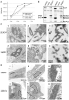
Figure 1
Figure 1. Infection of R06E cells with MARV and ZEBOV.
(A) VeroE6 or R06E cells were infected with MARV or ZEBOV with 0.5 TCID50/cell. Supernatants were collected at 1, 2, 3 and 7 days p.i. and used for TCID50 assays. (B) Supernatant from day 7 was concentrated via ultracentrifugation and viral particles were analyzed by Coomassie staining for protein composition. (C) R06E cells were infected with MARV and ZEBOV at a high MOI, fixed and inactivated at day 3 p.i.. Cells were dehydrated and embedded in Epon prior to ultrathin sectioning. Analysis by transmission electron microscopy showed viral inclusions in the perinuclear region (1, 2) and mature viral particles (3, 4). MARV and ZEBOV particles were purified via ultracentrifugation through a 20% sucrose cushion, fixed with 4% paraformaldehyde, negatively stained and analyzed by electron microscopy (5, 6). (D) VeroE6 and R06E cells were infected with MARV and ZEBOV at a high MOI, harvested at 48 h p.i. and treated as described under B. Analysis by transmission electron microscopy showed viral inclusions (broken lines) in the perinuclear region of VeroE6 (1, 2) and R06E (3, 4) cells at low magnification. Higher magnification pictures of viral inclusions in R06E cells are shown under 5 and 6.
Figure 2
Figure 2. Expression of viral proteins in different cell lines.
(A) 4×10⁵ HUH7, VeroE6 or R06E cells were infected with 0.1 TCID50/cell MARV or ZEBOV. Cell lysates were prepared at 48 and 72 h p.i., subjected to Western blot analysis to detect cellular tubulin and VP40 of MARV and ZEBOV using mouse monoclonal antibodies. The total amount of cellular proteins in the samples was quantified by separating cell lysates on SDS PAGE, which were then stained with Coomassie Blue. Using the Odyssey Infrared Imaging Application Software, the protein signals were quantified. VP40 levels normalized to total cell protein are shown in (B).
Figure 3
Figure 3. MARV-specific in vitro assays.
(A) Minigenome assay. Different cell lines were transfected with all of the plasmids necessary for replication and transcription of a MARV (3M–5M) minigenome. Relative light units (RLU) represent the efficiency of replication and transcription of the minigenome (upper panel). This experiment was performed in triplicate and standard deviations are shown. Asterisks indicate statistically significant differences (*** P-value≤0.002) RLUs shown in the upper panel were normalized to the transfection efficiency of the cells, as analyzed by a GFP-reporter construct. (lower panel) (B) iVLP assay with pretransfected indicator cells. HEK293 or R06E cells were transfected with all of the plasmids necessary to produce MARV iVLPs (producer cells, pc). Supernatants were collected 72 h p.t. and used to infect new indicator cells (HUH7 or R06E cells) pretransfected with all of the plasmids necessary for replication and transcription (indicator cells, ic). Three days p.i. the luciferase activity in the ic was determined, reflecting iVLP formation, budding, iVLP entry, minigenome delivery and secondary transcription. (C) iVLP assay with naïve indicator cells. R06E cells were transfected with all of the plasmids necessary to produce iVLPs (pc). Supernatant was collected 72 h p.t. and used to infect R06E cells that were not pretransfected. Luciferase activity in the indicator cells was determined 48 or 72 h p.i., reflecting iVLP formation, budding, iVLP entry, minigenome delivery and primary transcription of minigenomes. This experiment was performed in triplicate and standard deviations are shown.
Figure 4
Figure 4. Rescue of recombinant MARV from R06E cells.
R06E cells were transfected with all the plasmids sufficient for replication and transcription, a T7 polymerase construct and a T7-driven full-length cDNA construct of MARV (clone #16). (A) CPE formation was monitored at day 7 post transfection (p.t.). (B) Supernatant of transfected cells collected on day 7 was used to infect fresh VeroE6 cells (passage 1, p1). CPE formation was monitored 8 days post infection. (C) Supernatant of p1 was collected on day 8 p.i. and viral RNA was extracted. Glycoprotein gene-specific RT-PCR and subsequent restriction of the DNA with KpnI, a restriction site present only in the wild type genome, revealed the rescue of recombinant virus. (D) On day 8 p.i. supernatant and cells were lysed and subjected to Western blot analysis using monoclonal antibodies to detect the MARV proteins NP and VP40. * unknown cellular protein.

Similar articles

Cited by

References

    1. Bessa TA, Spichler A, Chapola EG, Husch AC, de Almeida MF, et al. The contribution of bats to leptospirosis transmission in Sao Paulo City, Brazil. Am J Trop Med Hyg. 2010;82:315–317. - PMC - PubMed
    1. Calisher CH, Childs JE, Field HE, Holmes KV, Schountz T. Bats: important reservoir hosts of emerging viruses. Clin Microbiol Rev. 2006;19:531–545. - PMC - PubMed
    1. Drexler JF, Corman VM, Gloza-Rausch F, Seebens A, Annan A, et al. Henipavirus RNA in African bats. PLoS One. 2009;4:e6367. - PMC - PubMed
    1. Halpin K, Young PL, Field HE, Mackenzie JS. Isolation of Hendra virus from pteropid bats: a natural reservoir of Hendra virus. J Gen Virol. 2000;81:1927–1932. - PubMed
    1. Lau SK, Woo PC, Li KS, Huang Y, Tsoi HW, et al. Severe acute respiratory syndrome coronavirus-like virus in Chinese horseshoe bats. Proc Natl Acad Sci U S A. 2005;102:14040–14045. - PMC - PubMed

Publication types