. 2010 Aug 24;5(8):e12369.
doi: 10.1371/journal.pone.0012369.

Dcas supports cell polarization and cell-cell adhesion complexes in development

Affiliations

Dcas supports cell polarization and cell-cell adhesion complexes in development

Nadezhda Tikhmyanova et al. PLoS One. .

Abstract

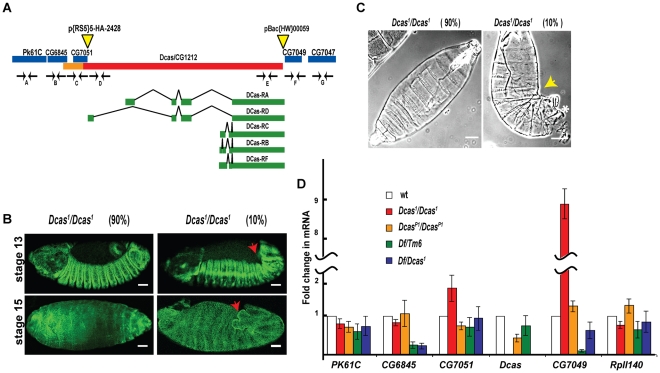
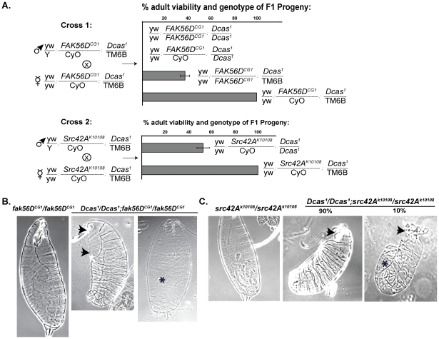
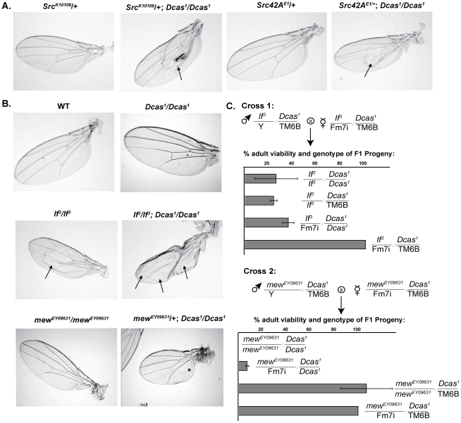
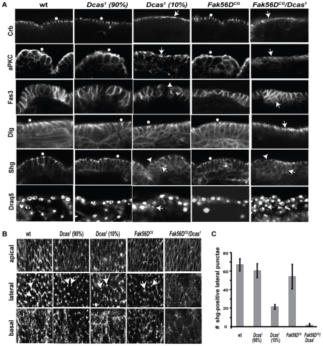
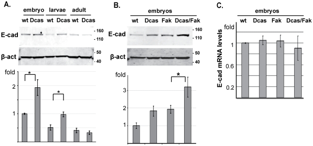
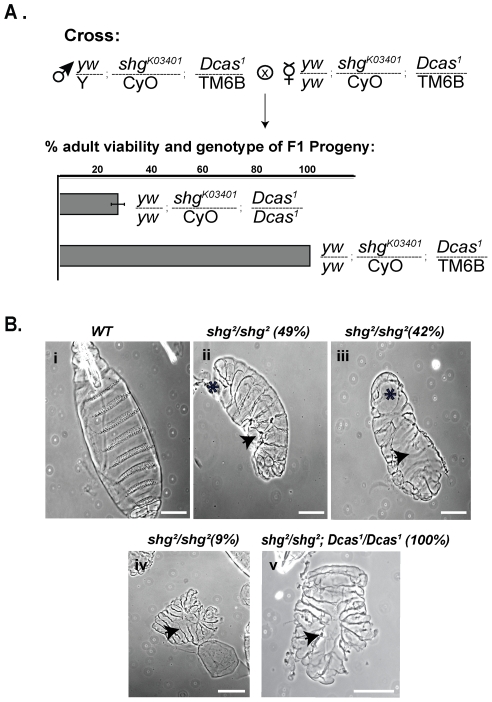
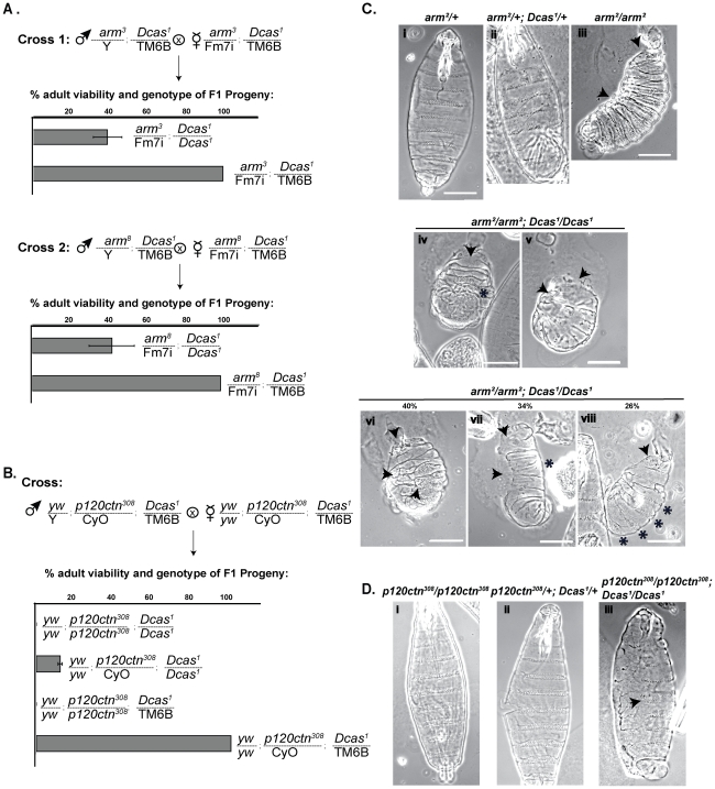
Mammalian Cas proteins regulate cell migration, division and survival, and are often deregulated in cancer. However, the presence of four paralogous Cas family members in mammals (BCAR1/p130Cas, EFS/Sin1, NEDD9/HEF1/Cas-L, and CASS4/HEPL) has limited their analysis in development. We deleted the single Drosophila Cas gene, Dcas, to probe the developmental function of Dcas. Loss of Dcas had limited effect on embryonal development. However, we found that Dcas is an important modulator of the severity of the developmental phenotypes of mutations affecting integrins (If and mew) and their downstream effectors Fak56D or Src42A. Strikingly, embryonic lethal Fak56D-Dcas double mutant embryos had extensive cell polarity defects, including mislocalization and reduced expression of E-cadherin. Further genetic analysis established that loss of Dcas modified the embryonal lethal phenotypes of embryos with mutations in E-cadherin (Shg) or its signaling partners p120- and beta-catenin (Arm). These results support an important role for Cas proteins in cell-cell adhesion signaling in development.


Conflict of interest statement

Competing Interests: The authors have declared that no competing interests exist.

Figures

Figure 1
Figure 1. Generation of the Dcas1 mutant stock.
A. Dcas (CG1212) locus, with coding region (red) and promoter (orange) indicated, with alternatively spliced variant transcripts indicated below the sequence (exons shown in green, introns as black lines). Flanking genes are shown in blue. Yellow triangles mark positions of P-element insertions p{RS5}5-HA-2428 and pBac{WH}00059 used to make the Dcas1 mutant. Position of primer pairs used in quantitative RT-PCR to confirm deletion of the Dcas gene (D, E) but not flanking genes (A, B, C, F, G) are indicated. B. Dorsal views of stage 13 and 16 Dcas1/Dcas1 embryos stained with Fas3, indicating phenotypes of 90% (left panel) and 10% (right panel) of mutant flies. The embryos are oriented anterior to the left. Red arrow indicates characteristic “fishtail” at posterior in the 10% of embryos with DC and GBR retraction defects. C. Cuticle preparations of Dcas1/Dcas1 mutant embryos; yellow arrow indicates DC and GBR defects, * indicates hole in posterior dorsal cuticle. Scale bar, 40 µm. D. Graph representing change in mRNA levels for indicated genes as measured by qRT-PCR analysis of cDNA prepared from wild type (white), Dcas1/Dcas1 (red) and DcasP1/DcasP1 (orange), Df(3L)Exel6083/+(green) and Df(3L)Exel6083/Dcas1 (blue). Bars represent standard error.
Figure 2
Figure 2. Genetic interactions of Dcas with Fak56D and Src42A.
A. Represent examples of a genetic cross of two double heterozygous parents (Fak56DCG1 and Dcas1 in Cross 1 or SrcK10108 and Dcas1 in Cross 2) to allow analysis of the viability of resulting progeny. Each row in the graph represents percentage of viable progeny of indicated genotype. Total number of expected progeny was calculated from the number of phenotypically viable double balanced adult heterozygotes Fak56CG1/CyO; Dcas1/TM6B, Ubx, y+ in Cross 1 and SrcK10108/CyO; Dcas1/TM6B, Ubx, y+ in Cross 2, which we considered 100% viable. The percentage of viability for the remaining progeny was calculated in agreement with Mendel's law of independent assortment for two alleles. B. Cuticle preparations of stage 16 Fak56DCG1/Fak56DCG1 and Fak56DCG1/Fak56DCG1;Dcas1/Dcas1 embryos transitioning to 1st instar larvae, viewed laterally, ventral side to right. Arrows indicate holes in head and dorsal cuticle, * indicates missing and/or fused denticle belts. C Cuticle preparations of stage 16 Src42Ak10108/Src42Ak10108 and Src42Ak10108/Src42Ak10108;Dcas1/Dcas1 embryos. Arrows indicates holes in head; * indicates GBR defect associated with incomplete DC.
Figure 3
Figure 3. Absence of Dcas induces wing defects in Src42A, If, and mew-deficient flies.
A. Dcas1/Dcas1 genotype induces a blister (arrows) phenotype in Src42A heterozygous mutant flies. B. Arrows point to typical wing blisters in wings of the flies of the indicated genotypes. C. Representative genetic cross of two double heterozygous parents (If3 and Dcas1; mewEY09631 and Dcas1) to allow analysis of the viability of resulting progeny. Each row in the graph represents percentage of viable progeny of indicated genotype. Only female progeny were analyzed.
Figure 4
Figure 4. Cell polarity consequences of mutations in Dcas and Fak56.
A. Immunofluorescence of epithelial cells of stage 15 embryos with indicated homozygous mutant genotypes, visualized with antibodies to Crb, aPKC, Fas3, Dlg, and Shg, as indicated. Arrows and arrowheads indicate defects in the localization of apico-basal polarity determinants or morphology changes in mutants, while asterisks indicate wild type appearance for each marker. In Crb panels, arrows point to a apically diffused localization of Crb in Dcas1 (10%) embryos, and reduced and diffuse localization of Crb in homozygotic Dcas1/Fak56CG1 embryos. In aPKC panels, arrows indicate abnormally punctate localization of Crb in Dcas1 (10%) embryos and diminished and diffuse localization of Crb in homozygotic Dcas1/Fak56CG1. The abnormally punctate and apical localization of Dlg is indicated with an arrow in homozygous Dcas1/Fak56CG1 embryos. In Shg panels, arrowheads point to cell junctions with reduced visibility of lateral punctae, and arrows indicate the increased cytoplasmic localization of Shg in Dcas1 (10%) and Dcas1/Fak56CG1 embryos. In Fas3, arrowhead points to a rounded cell within the epithelial layer in a Dcas1 (10%) embryo stained with Fas3. Multilayering of cells in the epithelium of Dcas1/Fak56CG1 and/or Dcas1 (10%) embryos is apparent in embryos stained with Fas3 or Draq5. B. Immunofluorescence with antibody to Shg visualizing apical, lateral, and basal z-series of stage 15 embryonal epithelial cells from flies with indicated genotypes. Z-sections were taken starting from the apical surface, with increments of 0.1 µm. Lateral images shown here reflect the 5th section (0.5 µm) and basal reflects the 10th section (1 µm) down from the apical surface. Lateral punctae are marked with arrows. C. Quantification of punctate E-cadherin-positive lateral junctions in flies of indicated homozygotic genotypes, per 35 µm2. More than 6 embryos in 3 independent experiments were analyzed.
Figure 5
Figure 5. Dcas negatively regulates expression of E-cadherin.
A. Western analysis of lysates prepared from wt or Dcas1/Dcas1 (Dcas) stage 13–16 embryos, 1st–2nd instar larvae, or adult flies with antibody to DE-cadherin. β-actin was used as loading control. Graph below compares E-cadherin normalized β-actin based on results of 3 independent experiments; *, P = 0.003 B. Western analysis of lysates from wt, Dcas1/Dcas1 (Dcas), FAK56DCG1/FAK56DCG1 (fak) and Dcas1/Dcas1; Fak56DCG1/Fak56DCG1 (Dcas/fak). Graph as in A, *, P = 0.005. C. Expression levels of E-cadherin mRNA in stage 13–16 embryos of the indicated genotypes, as established by RT-PCR. Differences are not statistically significant.
Figure 6
Figure 6. Genetic interactions of Dcas with shg.
A. Representative genetic cross of two double heterozygous parents (shgK03401 and Dcas1) to allow analysis of the viability of resulting progeny. Each row in the graph represents percentage of viable progeny of indicated genotype. B. Cuticle preparations of WT, shg2/shg2 and Dcas1/Dcas1;shg2/shg2 stage 16 embryos, viewed ventrally (panels i and iii), laterally (panel ii) and dorsally (panels iv–v). * indicates defects in head and ventral cuticle formation, respectively, arrows point to holes in ventral and dorsal cuticle. Genotypes and percentages of cuticles with indicated phenotypes are marked on top. Scale bar, 100 µm.
Figure 7
Figure 7. Genetic interactions of Dcas with arm, shg and p120ctn.
A, B. Representative genetic cross of two double heterozygous parents (arm3 and Dcas1, arm8 and Dcas1, p120ctn308 and Dcas1) to allow analysis of the viability of resulting progeny. Each row in the graph represents percentage of viable progeny of indicated genotype. C. Cuticle preparations of stage 16 embryos of the indicated genotypes. Panels iii and v–viii, lateral view; panels i, ii, and iv, ventral view. Arrows indicate holes in head and/or dorsal cuticle, and GBR defect; * indicates fused denticles in panel iv, lack of ventral denticles in panel vii, and well-separated ventral denticle belts in panel viii. Percentages of embryos with fused, absent and well-separated denticles are shown above in panels iv-vi. D. Cuticle preparations of stage 16 embryos of the indicated genotypes. Arrow indicates fused ventral denticle belts.

Similar articles

Cited by

References

    1. Defilippi P, Di Stefano P, Cabodi S. p130Cas: a versatile scaffold in signaling networks. Trends Cell Biol. 2006;16:257–263. - PubMed
    1. Singh M, Cowell L, Seo S, O'Neill G, Golemis E. Molecular basis for HEF1/NEDD9/Cas-L action as a multifunctional co-ordinator of invasion, apoptosis and cell cycle. Cell Biochem Biophys. 2007;48:54–72. - PMC - PubMed
    1. Tikhmyanova N, Little JL, Golemis EA. CAS proteins in normal and pathological cell growth control. Cell Mol Life Sci 2009 - PMC - PubMed
    1. Sakai R, Iwamatsu A, Hirano N, Ogawa S, Tanaka T, et al. A novel signaling molecule, p130, forms stable complexes in vivo with v-Crk and v-Src in a tyrosine phosphorylation-dependent manner. EMBO J. 1994;13:3748–3756. - PMC - PubMed
    1. Law SF, Estojak J, Wang B, Mysliwiec T, Kruh GD, et al. Human Enhancer of Filamentation 1 (HEF1), a novel p130Cas-like docking protein, associates with FAK, and induces pseudohyphal growth in yeast. Mol Cell Biol. 1996;16:3327–3337. - PMC - PubMed

Publication types

MeSH terms

LinkOut - more resources