. 2010 Aug 24;5(8):e12387.
doi: 10.1371/journal.pone.0012387.

GFAP-driven GFP expression in activated mouse Müller glial cells aligning retinal blood vessels following intravitreal injection of AAV2/6 vectors

Affiliations

GFAP-driven GFP expression in activated mouse Müller glial cells aligning retinal blood vessels following intravitreal injection of AAV2/6 vectors

Wendy M Aartsen et al. PLoS One. .

Abstract

Background: Müller cell gliosis occurs in various retinal pathologies regardless of the underlying cellular defect. Because activated Müller glial cells span the entire retina and align areas of injury, they are ideal targets for therapeutic strategies, including gene therapy.

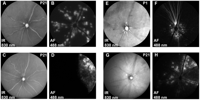
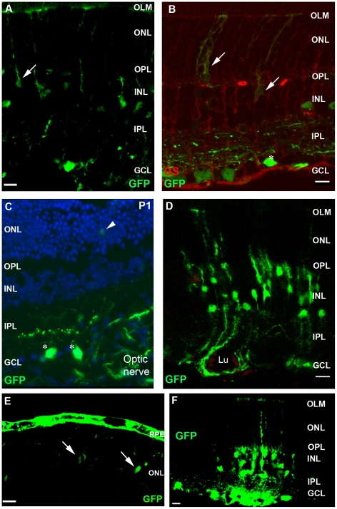
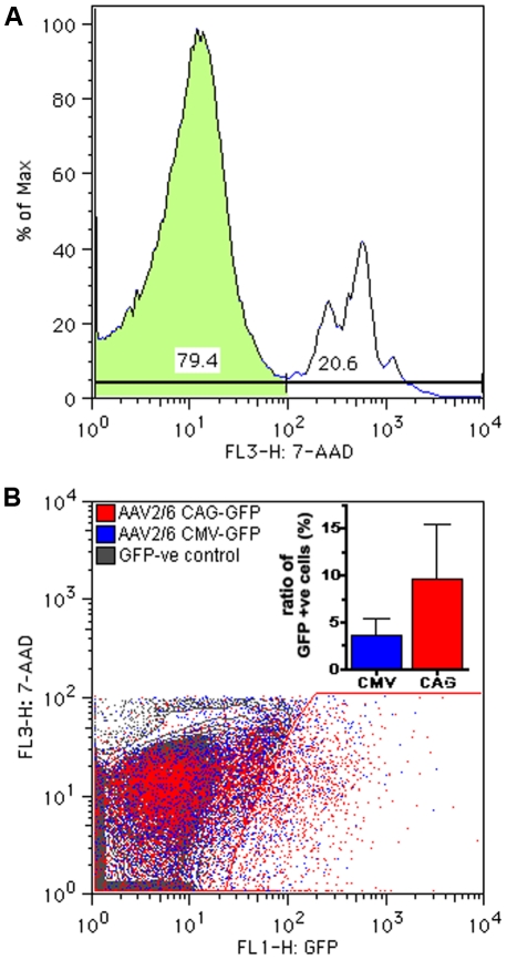
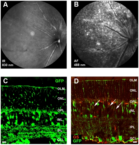
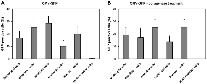
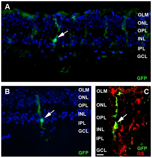
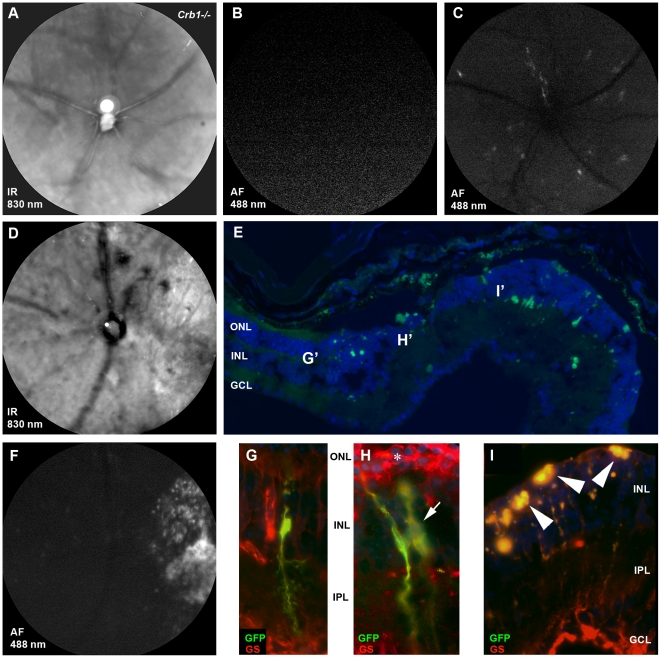
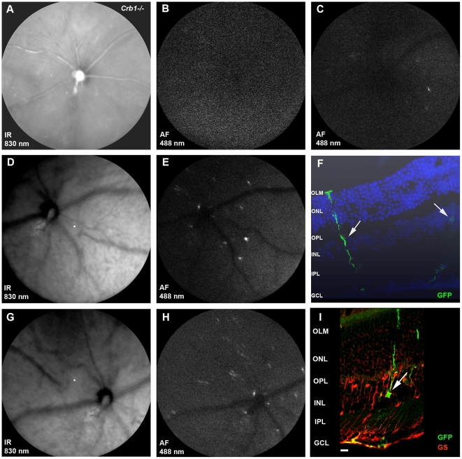
Methodology/principal findings: We used adeno-associated viral AAV2/6 vectors to transduce mouse retinas. The transduction pattern of AAV2/6 was investigated by studying expression of the green fluorescent protein (GFP) transgene using scanning-laser ophthalmoscopy and immuno-histochemistry. AAV2/6 vectors transduced mouse Müller glial cells aligning the retinal blood vessels. However, the transduction capacity was hindered by the inner limiting membrane (ILM) and besides Müller glial cells, several other inner retinal cell types were transduced. To obtain Müller glial cell-specific transgene expression, the cytomegalovirus (CMV) promoter was replaced by the glial fibrillary acidic protein (GFAP) promoter. Specificity and activation of the GFAP promoter was tested in a mouse model for retinal gliosis. Mice deficient for Crumbs homologue 1 (CRB1) develop gliosis after light exposure. Light exposure of Crb1(-/-) retinas transduced with AAV2/6-GFAP-GFP induced GFP expression restricted to activated Müller glial cells aligning retinal blood vessels.

Conclusions/significance: Our experiments indicate that AAV2 vectors carrying the GFAP promoter are a promising tool for specific expression of transgenes in activated glial cells.


Conflict of interest statement

Competing Interests: The authors have declared that no competing interests exist.

Figures

Figure 1
Figure 1. SLO of AAV2/6-CMV-GFP transduced wild-type mouse retinas 3 weeks post injection.
SLO analysis was performed at 830 (IR) and 488 (AF) nm on all animals and representative examples are shown. After intravitreal injection of AAV2/6-CMV-GFP at postnatal day 21 (n = 6), the integrity of the retina appeared normal (A) and the fluorescent signal for GFP was detected along the major retinal blood vessels (n = 6) (B). After subretinal injection with AAV2/6-CMV-GFP at postnatal day 21 (n = 6), retinal integrity was not affected (C), while the GFP expression was detected at a restricted part of the retina (D). Intravitreal injection of AAV2/6-CMV-GFP at postnatal day 1 (n = 5) did no major damage to the retina (E) and GFP expression was concentrated at the center of the retina near the optic nerve (F). Intravitreal injection of AAV2/6-CAG-GFP at postnatal day 21 (n = 5) did not harm the retinal integrity (G), while GFP expression was predominantly found along retinal blood vessels (H), comparable to AAV2/6-CMV-GFP.
Figure 2
Figure 2. Immunohistochemistry on AAV2/6-CMV-GFP transduced wild-type mouse retinas 3 weeks post injection.
Examples of transduced cell types after intravitreal injection of AAV2/6-CMV-GFP at postnatal day 21 (A) and confirmed by colocalization of GFP (green) and labeling with the Müller glia cell marker glutamine synthetase (red) (B). Transduction of mainly ganglion cells near the optic nerve when AAV2/6-CMV-GFP was injected at postnatal day 1 (C). Müller glial cells in the close proximity of a blood vessel (D). GFP expression in RPE and photoreceptors cells after injecting AAV2/6-CMV-GFP subretinally (P21, E). Transduced cell types found after intravitreal injection of AAV2/6-CAG-GFP at postnatal day 21 (F). Symbols/abbreviations used in panels: Arrow; Müller glial cells, Asterisks; ganglion cells, Arrow head; photoreceptor cells, ONL; outer nuclear layer, OPL; outer plexiform layer, INL; inner nuclear layer, IPL; inner plexiform layer, GCL; ganglion cell layer. Lu; vessel lumen. Scale bars represent 10 µm.
Figure 3
Figure 3. Flow cytometric analysis of AAV2/6-CMV-GFP vs AAV2/6-CAG-GFP transduced retinas.
Representative example of 7-AAD staining; the negative (live) population is used for further analysis (A). A representative comparison of retinal cells from eyes transduced via intravitreal injection of either AAV2/6-CMV-GFP (blue) or AAV2/6-CAG-GFP (red) resulted in a population of GFP-positive cells. The left edge of the gate for GFP-positive cells was set using non-transduced retinal cells (gray) (B). The inset shows the mean (+SEM) of the fraction of the live cells that are GFP-positive in CAG-GFP compared to CMV-GFP eyes (n = 5). These measurements show no statistically significant differences (p>0.6, Mann-Whitney test).
Figure 4
Figure 4. The ILM forms a barrier for efficient Müller glial cell transduction by AAV2/6-CMV-GFP.
Representative images of SLO at 830 nm (A) and 488 nm (B) and of fluorescence microscopy for GFP expression (C and D) are shown for retinas after intravitreal injection of AA2/6-CMV-GFP in collagenase treated wild-type eyes (n = 4) at three weeks post injection. The integrity of the retinas appeared normal in most cases (A). GFP expression was detected throughout the whole retina (B). Sections of the retina reveal transduction of Müller glial cells, ganglion, amacrine, horizontal and bipolar cells (C). The cell type was confirmed by double labeling with the Müller glial cell marker glutamine synthetase (red) (D). Symbols/abbreviations used in panels: Arrow; Müller glial cells, Asterisks; ganglion cells, OLM; outer limiting membrane, ONL; outer nuclear layer, OPL; outer plexiform layer, INL; inner nuclear layer, IPL; inner plexiform layer, GCL; ganglion cell layer. Scale bars represent 10 µm.
Figure 5
Figure 5. Transduction profiles of AAV2/6-CMV-EGFP retinas with or without collagenase treatement.
Representative retinal slices from injected eyes were quantified for the number of each transduced cell type. Specific markers were used to determine the different cell types and to generate histograms comparing tropism profiles with (B) and without collagenase treatment (A). Transduction efficiencies were calculated based on the ratio of each cell type infected relative to the total number of EGFP-positive cells (n = 9). Error bars represent standard deviation among sample population.
Figure 6
Figure 6. Transduction of human cultured retinas with AAV2/6-CMV-GFP.
Retinas were prepared from donor eyes and transduced with AA2/6-CMV-GFP, by injection of 1 µl viral particle suspension under the ILM. Retinas were cultured for 7 days (n = 2), followed by fluorescence microscopic analysis of cryostat sections (10 µm). GFP-positive Müller glial cells (green signals) were found near the injection site (A) as well as further away (B). The cell type was confirmed by double labeling with the Müller glial cell marker glutamine synthetase (red) (C). Symbols/abbreviations used in panels: Arrow; Müller glial cells, OLM; outer limiting membrane, ONL; outer nuclear layer, OPL; outer plexiform layer, INL; inner nuclear layer, IPL; inner plexiform layer, GCL; ganglion cell layer.
Figure 7
Figure 7. Light-induced GFP expression in AAV2/6-GFAP-GFP transduced Crb1−/− Müller glial cells.
Crb1−/− mice, three weeks of age, were intravitreally injected with AAV2/6-GFAP-GFP (8×10⁹ genome copies; n = 3). At 21 days after injection, before light exposure, retinal degeneration and fluorescence were analyzed by SLO at 830 nm (A) and 488 nm (B), respectively. 10 weeks after transduction the mice were exposed to white light (3000 lux) for 72 hours. GFAP-driven GFP expression was detected by SLO in Crb1−/− mice (n = 7) (C). One out of seven Crb1−/− animals showed severe retinal degeneration on SLO analysis as reported before , (D). The retinas showed extensive fluorescence in one quadrant of the retina (F). In the severely affected retina (E), most GFP and glutamine synthetase positive Müller glial cells were found at the border of the area of ONL degeneration (G and H). Slices adjacent to the one showing the overview in E were subjected to immunohistochemistry with antibodies against glutamine synthetase. G', H' and I' are the positions of the figures G, H and I taken from adjacent slices to the overview shown in E. The asterisk in (H) points at the abnormal structure of the activated Müller glial cells and glial scar formation. In the middle of the degenerative area, the ONL is completely lost (E). In this area, highly auto-fluorescent patches of cell debris, indicated by arrowheads (I), accounted for most of the fluorescent signal found on SLO. Symbols/abbreviations used in panels: Arrow; Müller glial cells, ONL; outer nuclear layer, INL; inner nuclear layer, IPL; inner plexiform layer, GCL; ganglion cell layer.
Figure 8
Figure 8. CNTF-induced GFP expression in AAV2/6-GFAP-GFP transduced Müller glial cells.
3-week-old Crb1−/− mice were intravitreally injected with AAV2/6-GFAP-GFP (8×10⁹ genome copies; n = 3). Three weeks after injection, the integrity of the retina appeared normal on SLO (A). No GFP signal was observed (B). At 28 days after transduction, eyes were re-injected intravitreally with CNTF. GFAP-driven GFP expression was detected by SLO at one day (C) and four days (E and H) after administration of CNTF. Secondary injections did not affect the retinal integrity (D and G). Activated Müller glial cells expressing GFP were found on sections (10 µm) of the CNTF challenged eyes (F) and immuno-histochemistry confirmed the cell type as colocalization with glutamine synthetase was demonstrated (I). Symbols/abbreviations used in panels: Arrow; Müller glial cells OLM; outer limiting membrane, ONL; outer nuclear layer, OPL; outer plexiform layer, INL; inner nuclear layer, IPL; inner plexiform layer, GCL; ganglion cell layer.

References

    1. Bringmann A, Pannicke T, Grosche J, Francke M, Wiedemann P, et al. Muller cells in the healthy and diseased retina. Prog Retin Eye Res. 2006;25:397–424. - PubMed
    1. Dyer MA, Cepko CL. Control of Muller glial cell proliferation and activation following retinal injury. Nat Neurosci. 2000;3:873–880. - PubMed
    1. Goldstein IM, Ostwald P, Roth S. Nitric oxide: a review of its role in retinal function and disease. Vision Res. 1996;36:2979–2994. - PubMed
    1. Witmer AN, Vrensen GF, van Noorden CJ, Schlingemann RO. Vascular endothelial growth factors and angiogenesis in eye disease. Prog Retin Eye Res. 2003;22:1–29. - PubMed
    1. Bainbridge JW, Tan MH, Ali RR. Gene therapy progress and prospects: the eye. Gene Ther. 2006;13:1191–1197. - PubMed

Publication types

Substances