. 2010 Aug 24;8(8):e1000465.
doi: 10.1371/journal.pbio.1000465.

IL-6 and IL-10 anti-inflammatory activity links exercise to hypothalamic insulin and leptin sensitivity through IKKbeta and ER stress inhibition

Affiliations

IL-6 and IL-10 anti-inflammatory activity links exercise to hypothalamic insulin and leptin sensitivity through IKKbeta and ER stress inhibition

Eduardo R Ropelle et al. PLoS Biol. .

Retraction in

Abstract

Overnutrition caused by overeating is associated with insulin and leptin resistance through IKKbeta activation and endoplasmic reticulum (ER) stress in the hypothalamus. Here we show that physical exercise suppresses hyperphagia and associated hypothalamic IKKbeta/NF-kappaB activation by a mechanism dependent upon the pro-inflammatory cytokine interleukin (IL)-6. The disruption of hypothalamic-specific IL-6 action blocked the beneficial effects of exercise on the re-balance of food intake and insulin and leptin resistance. This molecular mechanism, mediated by physical activity, involves the anti-inflammatory protein IL-10, a core inhibitor of IKKbeta/NF-kappaB signaling and ER stress. We report that exercise and recombinant IL-6 requires IL-10 expression to suppress hyperphagia-related obesity. Moreover, in contrast to control mice, exercise failed to reverse the pharmacological activation of IKKbeta and ER stress in C3H/HeJ mice deficient in hypothalamic IL-6 and IL-10 signaling. Hence, inflammatory signaling in the hypothalamus links beneficial physiological effects of exercise to the central action of insulin and leptin.


Conflict of interest statement

The authors have declared that no competing interests exist.

Figures
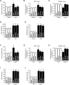
Figure 1
Figure 1. Exercise induces appetite-suppressive actions in different models of obesity.
(A) 12 h of food intake (kcal) in lean and diet-induced obesity (DIO) Wistar rats under resting conditions or after swimming exercise (SW Exe) or treadmill running (TR Exe) (n = 20–35 animals per group). Rats were fasted during 9 h and the hypothalamic levels (B) NPY and (C) POMC mRNA were examined using real time PCR assay. (D) Body weight, (E) epididymal fat pad weight, (F) 12-h food intake of leptin-deficient mice (Leptob/ob) and respective wild type group. (G) NPY and (H) POMC mRNA were examined using real time PCR assay. (I) Body weight and (J) epididymal fat pad weight of wild type and leptin-deficient mice under resting conditions or immediately after the exercise protocols (n = 10 animals per group). Data are the means ± SEM. # p<0.05 versus respective lean group at rest; * p<0.05 versus respective obese group at rest. Lean animals (white bars) and obese animals (black bars).
Figure 2
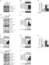
Figure 2. Hypothalamic insulin and leptin signaling after exercise.
Western blots showing hypothalamic lysates from Wistar rats; (A) Hypothalamic IRβ, IRS-1, IRS-2, and Akt phosphorylation, (B) Hypothalamic Foxo1 phosphorylation. (C) 12-h food intake (kcal) after intrahypothalamic infusion of insulin in lean and diet-induced obesity (DIO) Wistar rats under resting conditions or after exercise (n = 6–8 animals per group). Western blots of five independent experiments showing hypothalamic lysates from Wistar rats; (D) Insulin-induced IRβ, IRS-1, IRS-2, Akt, and Foxo1 phosphorylation in the hypothalamus. (E) Subcellular fractionation was performed to evaluate the nuclear Foxo1 expression in the hypothalamus of lean and obese rats at 30 min after insulin infusion. (F) Hypothalamic Jak-2 and (G) STAT-3 tyrosine phosphorylation. (H) 12-h food intake (kcal) after intrahypothalamic infusion of leptin (n = 6–8 animals per group). Western blots showing hypothalamic lysates from Wistar rats; (I) Leptin-induced Jak2, IRS-1, IRS-2, and STAT3 tyrosine phosphorylation in the hypothalamus. (J) Subcellular fractionation was performed to evaluate the nuclear STAT3 expression in the hypothalamic cells of lean and obese rats 30 min after leptin infusion. Data are the means ± SEM. # p<0.05 versus respective lean group at rest; * p<0.05 versus obese group at rest. Lean animals (white bars) and obese animals (black bars). The Figure 2F IB: pJak2Tyr1007/8 panel is excluded from the article's copyright license. See the accompanying retraction notice for more information.
Figure 3
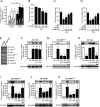
Figure 3. Anti-hyperphagic response mediated by IL-6.
(A) IL-6 mRNA in the hypothalamus of lean or diet-induced obesity (DIO) rats under resting conditions and lean obese rats immediately after the swimming exercise (SW Exe) or treadmill running (TR Exe). (B) 12 h of food intake in obese rats under resting conditions following intrahypothalamic infusion of different doses of recombinant IL-6. Counter-regulatory effects of anti-IL-6 antibody on food intake in exercised obese rats after (C) insulin or (D) leptin infusion. Western blots of five independent experiments showing hypothalamic lysates from Wistar rats; (E) Expression and activity of protein involved in the inflammatory signaling or ER stress in control animals at rest condition or after acute exercise (F) TLR4 expression, (G) IKKβ phosphorylation, (H) IκBα expression, (I) PERK phosphorylation, (J) CHOP expression, and (K) IRS-1Ser307 phosphorylation from lean, obese, obese injected with recombinant IL-6, obese after exercise, and obese pretreated with anti-IL-6 antibody before the exercise protocol. Data are the means ± SEM. # p<0.05 versus lean group; * p<0.05 versus obese group at rest; ¥ p<0.05 versus respective exercised control rats; ** p<0.01 versus stimulated obese group at rest; § p<0.05 versus obese group injected with recombinant IL-6 and exercised obese rats (n = 8–10 animals per group). Swimming Exercise (SW Exe) or Treadmill Running (TR Exe). Lean animals (white bars) and obese animals (black bars).
Figure 4
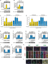
Figure 4. IL-6R localization in the hypothalamus of rats.
(A) Immunohistochemistry was performed in the hypothalamic tissue of control rats, using IL-6 receptor (IL-6R)-specific antibody (green) and DAPI (blue), with 50× magnification. (B) Positive cells were quantified in different hypothalamic nuclei, § p<0.05 versus the other nuclei. (C) In situ hybridization showing the co-localization of IL-6R (red) with POMC, NPY, and AgRP (green) neuropeptides in the hypothalamus of control rats. Head arrows show neurons and arrows show endothelial cells using 20× and 63× magnification. (D) The dissection of hypothalamic arcuate nucleus of lean and obese rats was obtained as described in Experimental Procedures to evaluate the mRNA of POMC, NPY, and AgRP, using the real time PCR. Data are the means ± SEM. # p<0.05 versus respective control group at rest; * p<0.05 versus obese rats at rest. Lean animals (yellow bars) and obese (blue bars). (E) Confocal microscopy was performed to evaluate the co-localization of IL-6R (green) and IKKβ, PERK, and IRS-1 (red) in the arcuate nuclei of obese rats, with 200× magnification (scale bar, 20 µm).
Figure 5
Figure 5. IL-6 reversed pharmacological endoplasmatic reticulum stress induction in the hypothalamus.
(A) 12 h of food intake in lean rats after thapsigargin infusion (3 µg). (B) Anorexigenic effects of insulin in the hypothalamus of lean rats pretreated with thapsigargin. (C) Anorexigenic effects of leptin in the hypothalamus of lean rats pretreated with thapsigargin. Western blots showing hypothalamic lysates from Wistar rats; (D) IKKβ, (E) PERK, and (F) IRS-1Ser307 phosphorylation from lean rats pretreated with thapsigargin. (G) Insulin-induced Akt serine phosphorylation, (H) leptin-induced STAT3 tyrosine phosphorylation in the hypothalamus of lean animals pretreated with thapsigargin, and (I) basal levels of Akt and STAT3 phosphorylation. Data are the means ± SEM. # p<0.05 versus DMSO group; * p<0.05 versus lean plus thapsigargin; § p<0.05 versus thapsigargin plus recombinant IL-6 or thapsigargin plus exercised (n = 8–10 animals per group).
Figure 6
Figure 6. Role of hypothalamic IL-10 in the control of energy intake during obesity.
Western blots showing hypothalamic lysates from Wistar rats; (A) IL-1ra and (B) IL-10 expression in the hypothalamus. (C) IL-10 mRNA in the hypothalamus was examined using real time PCR assay. (D) 12 h food intake (kcal) in obese rats under resting conditions after intrahypothalamic infusion of different doses of recombinant IL-10. Western blots showing hypothalamic lysates from Wistar rats; (E) IL-10 expression after ASO IL-10 treatment in obese animals. (F) Intrahypothalamic treatment with ASO IL-10 blocked the anorexigenic response mediated by (F) insulin and (G) leptin in exercised obese animals or obese animals at rest injected with recombinant IL-6. Western blots showing hypothalamic lysates from Wistar rats; (H) IKKβ, (I) PERK, and (J) IRS-1Ser307 phosphorylation after ASO IL-10 treatment or after acute recombinant IL-10 infusion. (K) Insulin-induced Akt serine phosphorylation and (L) leptin-induced STAT3 tyrosine phosphorylation in the hypothalamus after ASO IL-10 treatment or after acute recombinant IL-10 infusion. (M) Basal levels of Akt serine phosphorylation and (N) STAT3 tyrosine phosphorylation in the hypothalamus after ASO IL-10 treatment or after acute recombinant IL-10 infusion. Data are the means ± SEM. # p<0.05 versus chow group; * p<0.05 versus DIO; ¥ p<0.05 versus exercised control animals; n = 8–10 animals per group. Lean animals (white bars), obese animals (black bars), and exercised obese plus recombinant IL-10 (grey bars). SO, sense oligonucleotide; ASO, antisense oligonucleotide.
Figure 7
Figure 7. The central anti-inflammatory response mediated by exercise requires augmented hypothalamic levels of IL-6 and IL-10.
Western blots showing hypothalamic lysates from C3H/NeN and C3H/HeJ mice under resting conditions or after physical activity; (A) IL-6 and (B) IL-10 expression. Anorexigenic effects of insulin (C) or leptin (D) in C3H/NeN and C3H/HeJ mice under resting conditions, after thapsigargin, thapsigargin plus exercise, and thapsigargin plus recombinant IL-6 or IL-10. Western blots showing hypothalamic lysates from mice; (E) IL-10 expression at 2 h after intrahypothalamic injection of recombinant IL-6 (200 ng) in C3H/NeN and C3H/HeJ mice under resting conditions. (F) IKKβ, (G) PERK, and (H) IRS-1Ser307 phosphorylation and (I) Insulin-induced Akt serine phosphorylation and (J) leptin-induced STAT3 tyrosine phosphorylation in the hypothalamus of C3H/HeJ mice after intrahypothalamic infusion of DMSO, thapsigargin, thapsigargin plus exercise, and thapsigargin plus recombinant IL-6 or IL-10. Data are the means ± SEM. ** p<0.05 versus respective control group at rest; # p<0.05 versus respective control group non-stimulated or stimulated with DMSO; * p<0.05 versus thapsigargin; n = 5–6 animals per group. C3H/NeN (yellow bars) and C3H/HeJ (blue bars). (K) Co-localization of IL-10R (red) with NPY, AgRP, and POMC (green) was evaluated using in situ hybridization technique in the hypothalamus of lean rats, with 20× and 63× magnification. (L) Co-localization of IL-6R (green) and IL-10R (red) in the arcuate nuclei of lean rats, with 200× magnification (scale bar, 10 µm).
Figure 8
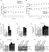
Figure 8. Effects of chronic exercise on food consumption, body weight, and IL-6 and IL-10 production.
Evaluation of (A) food intake (kcal) and (B) body weight in control and obese animals during chronic exercise protocol. Chow rest (black square), chow exercise (white square), DIO rest (black ball), and DIO exercise (white ball). (C) Body weight change between the 1st and 24th day. (D) Epididymal fat pad weight after chronic exercise, (E) IL-6 and (F) IL-10 mRNA levels in the hypothalamus of lean and obese rats at rest or after chronic exercise. Western blots showing hypothalamic lysates from lean and obese Wistar rats; (G) IKKβ phosphorylation and IκBα expression and (H) PERK phosphorylation and CHOP expression 1 and 24 d after the chronic exercise protocol. Data are the means ± SEM. * p<0.05 versus chow at rest; § p<0.05 versus DIO at rest; # p<0.05 versus chow group (rest); n = 8–10 animals per group. Lean animals (white bars) and obese animals (black bars).
Figure 9
Figure 9. Schematic diagrams of the proposed role of the hypothalamic anti-inflammatory response mediated by exercise.
(A) Overnutrition induces hypothalamic IKKβ activation and endoplasmatic reticulum stress, leading to central insulin and leptin resistance, hyperphagia, and obesity. (B) We propose that exercise increases the central anti-inflammatory response, increasing hypothalamic IL-6 and IL-10 expression. This phenomenon is crucial for reducing hypothalamic IKKβ activation and endoplasmatic reticulum stress and turn, restoring insulin and leptin signaling, and reorganizing the set point of nutritional balance.

Comment in

Similar articles

Cited by

References

    1. Carvalheira J. B, Ribeiro E. B, Araujo E. P, Guimaraes R. B, Telles M. M, et al. Selective impairment of insulin signalling in the hypothalamus of obese Zucker rats. Diabetologia. 2003;46:1629–1640. - PubMed
    1. Niswender K. D, Morton G. J, Stearns W. H, Rhodes C. J, Myers M. G, Jr, et al. Intracellular signalling. Key enzyme in leptin-induced anorexia. Nature. 2001;413:794–795. - PubMed
    1. Bruning J. C, Gautam D, Burks D. J, Gillette J, Schubert M, et al. Role of brain insulin receptor in control of body weight and reproduction. Science. 2000;289:2122–2125. - PubMed
    1. El-Haschimi K, Pierroz D. D, Hileman S. M, Bjorbaek C, Flier J. S. Two defects contribute to hypothalamic leptin resistance in mice with diet-induced obesity. J Clin Invest. 2000;105:1827–1832. - PMC - PubMed
    1. De Souza C. T, Araujo E. P, Bordin S, Ashimine R, Zollner R. L, et al. Consumption of a fat-rich diet activates a proinflammatory response and induces insulin resistance in the hypothalamus. Endocrinology. 2005;146:4192–4199. - PubMed

Publication types

MeSH terms