Oral N-acetyl-cysteine attenuates loss of dopaminergic terminals in alpha-synuclein overexpressing mice
- PMID: 20808797
- PMCID: PMC2925900
- DOI: 10.1371/journal.pone.0012333
Oral N-acetyl-cysteine attenuates loss of dopaminergic terminals in alpha-synuclein overexpressing mice
Abstract
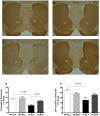
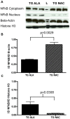
Levels of glutathione are lower in the substantia nigra (SN) early in Parkinson's disease (PD) and this may contribute to mitochondrial dysfunction and oxidative stress. Oxidative stress may increase the accumulation of toxic forms of alpha-synuclein (SNCA). We hypothesized that supplementation with n-acetylcysteine (NAC), a source of cysteine--the limiting amino acid in glutathione synthesis, would protect against alpha-synuclein toxicity. Transgenic mice overexpressing wild-type human alpha-synuclein drank water supplemented with NAC or control water supplemented with alanine from ages 6 weeks to 1 year. NAC increased SN levels of glutathione within 5-7 weeks of treatment; however, this increase was not sustained at 1 year. Despite the transient nature of the impact of NAC on brain glutathione, the loss of dopaminergic terminals at 1 year associated with SNCA overexpression was significantly attenuated by NAC supplementation, as measured by immunoreactivity for tyrosine hydroxylase in the striatum (p = 0.007; unpaired, two-tailed t-test), with a similar but nonsignificant trend for dopamine transporter (DAT) immunoreactivity. NAC significantly decreased the levels of human SNCA in the brains of PDGFb-SNCA transgenic mice compared to alanine treated transgenics. This was associated with a decrease in nuclear NFkappaB localization and an increase in cytoplasmic localization of NFkappaB in the NAC-treated transgenics. Overall, these results indicate that oral NAC supplementation decreases SNCA levels in brain and partially protects against loss of dopaminergic terminals associated with overexpression of alpha-synuclein in this model.
Conflict of interest statement
Figures


Similar articles
-
Mutant α-Synuclein Overexpression Induces Stressless Pacemaking in Vagal Motoneurons at Risk in Parkinson's Disease.J Neurosci. 2017 Jan 4;37(1):47-57. doi: 10.1523/JNEUROSCI.1079-16.2016. J Neurosci. 2017. PMID: 28053029 Free PMC article.
-
N-acetylcysteine increases dopamine release and prevents the deleterious effects of 6-OHDA on the expression of VMAT2, α-synuclein, and tyrosine hydroxylase.Neurol Res. 2024 May;46(5):406-415. doi: 10.1080/01616412.2024.2325312. Epub 2024 Mar 18. Neurol Res. 2024. PMID: 38498979
-
AAV1/2-induced overexpression of A53T-α-synuclein in the substantia nigra results in degeneration of the nigrostriatal system with Lewy-like pathology and motor impairment: a new mouse model for Parkinson's disease.Acta Neuropathol Commun. 2017 Feb 1;5(1):11. doi: 10.1186/s40478-017-0416-x. Acta Neuropathol Commun. 2017. PMID: 28143577 Free PMC article.
-
Reprint of: revisiting oxidative stress and mitochondrial dysfunction in the pathogenesis of Parkinson disease-resemblance to the effect of amphetamine drugs of abuse.Free Radic Biol Med. 2013 Sep;62:186-201. doi: 10.1016/j.freeradbiomed.2013.05.042. Epub 2013 Jun 3. Free Radic Biol Med. 2013. PMID: 23743292 Review.
-
Could a loss of alpha-synuclein function put dopaminergic neurons at risk?J Neurochem. 2004 Jun;89(6):1318-24. doi: 10.1111/j.1471-4159.2004.02423.x. J Neurochem. 2004. PMID: 15189334 Review.
Cited by
-
Glutathione Depletion and MicroRNA Dysregulation in Multiple System Atrophy: A Review.Int J Mol Sci. 2022 Dec 1;23(23):15076. doi: 10.3390/ijms232315076. Int J Mol Sci. 2022. PMID: 36499400 Free PMC article. Review.
-
Autophagy as an essential cellular antioxidant pathway in neurodegenerative disease.Redox Biol. 2013 Dec 25;2:82-90. doi: 10.1016/j.redox.2013.12.013. eCollection 2014. Redox Biol. 2013. PMID: 24494187 Free PMC article. Review.
-
N-Acetylcysteine breaks resistance to trastuzumab caused by MUC4 overexpression in human HER2 positive BC-bearing nude mice monitored by 89Zr-Trastuzumab and 18F-FDG PET imaging.Oncotarget. 2017 Apr 10;8(34):56185-56198. doi: 10.18632/oncotarget.17015. eCollection 2017 Aug 22. Oncotarget. 2017. PMID: 28915583 Free PMC article.
-
DNA damage and transcription stress cause ATP-mediated redesign of metabolism and potentiation of anti-oxidant buffering.Nat Commun. 2019 Oct 25;10(1):4887. doi: 10.1038/s41467-019-12640-5. Nat Commun. 2019. PMID: 31653834 Free PMC article.
-
α-synucleinopathy exerts sex-dimorphic effects on the multipurpose DNA repair/redox protein APE1 in mice and humans.Prog Neurobiol. 2022 Sep;216:102307. doi: 10.1016/j.pneurobio.2022.102307. Epub 2022 Jun 13. Prog Neurobiol. 2022. PMID: 35710046 Free PMC article.
References
-
- Thomas B, Beal MF. Parkinson's disease. Hum Mol Genet. 2007;16(R2):R183–194. - PubMed
-
- Schapira AH. Progress in Parkinson's disease. Eur J Neurol. 2008;15(1):1. - PubMed
-
- Schapira AH, et al. Mitochondrial function in Parkinson's disease. The Royal Kings and Queens Parkinson's Disease Research Group. Ann Neurol. 1992;32(Suppl):S116–24. - PubMed
-
- Greenamyre JT, Betarbet R, Sherer TB. The rotenone model of Parkinson's disease: genes, environment and mitochondria. Parkinsonism Relat Disord. 2003;9(Suppl 2):S59–64. - PubMed
-
- Langston JW. The etiology of Parkinson's disease with emphasis on the MPTP story. Neurology. 1996;47(6)(Suppl 3):S153–60. - PubMed
Publication types
MeSH terms
Substances
LinkOut - more resources
Full Text Sources
Other Literature Sources
Molecular Biology Databases
Miscellaneous

