. 2010 Aug 19;5(8):e12302.
doi: 10.1371/journal.pone.0012302.

A Toxoplasma MORN1 null mutant undergoes repeated divisions but is defective in basal assembly, apicoplast division and cytokinesis

Affiliations

A Toxoplasma MORN1 null mutant undergoes repeated divisions but is defective in basal assembly, apicoplast division and cytokinesis

Alexander Lorestani et al. PLoS One. .

Abstract

The membrane occupation and recognition nexus protein 1 (MORN1) is highly conserved among apicomplexan parasites and is associated with several structures that have a role in cell division. Here we dissected the role of MORN1 using the relatively simple budding process of Toxoplasma gondii as a model. Ablation of MORN1 in a conditional null mutant resulted in pronounced defects suggesting a central role for MORN1 in apicoplast segregation and in daughter cell budding. Lack of MORN1 resulted in double-headed parasites. These Janus-headed parasites form two complete apical complexes but fail to assemble a basal complex. Moreover, these parasites were capable of undergoing several more budding rounds resulting in the formation of up to 16-headed parasites conjoined at the basal end. Despite this segregation defect, the mother's cytoskeleton was completely disassembled in every budding round. Overall this argues that successful completion of the budding is not required for cell cycle progression. None of the known basal complex components, including a set of recently identified inner membrane complex (IMC) proteins, localized correctly in these multi-headed parasites. These data suggest that MORN1 is essential for assembly of the basal complex, and that lack of the basal complex abolishes the contractile capacity assigned to the basal complex late in daughter formation. Consistent with this hypothesis we observe that MORN1 mutants fail to efficiently constrict and divide the apicoplast. We used the null background provided by the mutant to dissect the function of subdomains of the MORN1 protein. This demonstrated that deletion of a single MORN domain already prevented the function of MORN1 whereas a critical role for the short linker between MORN domains 6 and 7 was identified. In conclusion, MORN1 is required for basal complex assembly and loss of MORN1 results in defects in apicoplast division and daughter segregation.


Conflict of interest statement

Competing Interests: The authors have declared that no competing interests exist.

Figures

Figure 1
Figure 1. Establishment and validation of a conditional MORN1 knock-out parasite.
(A–E) MORN1 expression levels assessed in pseudo-diploid parasites expressing an ATc conditional Myc2-tagged MORN1 allele driven by the Tet7sag1 promoter next to the endogenous MORN1 allele. (A) Total lysates harvested from uninduced (-ATc) and 24 hrs induced (+ATc) parasites were western blotted and probed with MORN1 and Myc antibodies as indicated. Arrows mark the Myc tagged MORN1; the arrowhead marks endogenous MORN1. (B–E). MORN1 (green) and Myc (red) expression under the same conditions as (A) assessed by immunofluorescence. (F) Time-course of MORN1 expression in the knock-out parasites at 0, 24, 48 and 96 hrs post ATc addition. (G–J) Plaque assays of the MORN1-KO parasites in absence of ATc (G), under ATc for 7 days (H) or 20 days (I) and for 7 days under ATc followed by 9 days without ATc (J). The absence of plaques in H and I indicates no viable parasites whereas the small plaque size and low numbers in panel J indicate aborted recovery of the phenotype. Additional recovery assays are shown in Supplementary Figure S2. A concentration of 1.0 µg/ml ATc was used throughout all experiments.
Figure 2
Figure 2. MORN1 knock-out tachyzoites display a lethal phenotype.
(A) Growth curves of the conditional MORN1 knock-out parasites with and without ATc induction displaying a decreased growth rate at 24 hrs post induction. Vacuole sizes of unsynchronized parasites were scored by fluorescence microscopy on fixed parasites using IMC3 antiserum; error bars denote standard deviations of three independent experiments. (B, C) Developmental progression analysis through the tachyzoite division cycle upon 18 hrs (B) and 24 hrs (C) of ATc induction. Developmental stages were defined using centrosome and daughter budding markers as indicated in (D–H). Cultures were unsynchronized and over 100 vacuoles per condition were counted in three independent experiments; error bars denote standard deviation. (D–H) Phenotype development was assessed using centrosome (α-centrin) and cytoskeleton scaffold (α-IMC1) markers as well as DAPI for the nuclear material. Stages were defined as indicated at the bottom right; numbers indicate the number of either centrosomes “C”, daughter buds “B” or nuclei “N”. Stages 2N2C and 2N4C were only detected in the ATc induced samples. Classes shown in (G) and (H) were differentiated for presence of buds as well, but this percentage was very low (1–2%) and is not included in the graphs.
Figure 3
Figure 3. MORN1-KO mutants display a multi-headed, division phenotype.
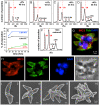
(A–F) DNA content was assessed by flow cytometry with or without ATc for time periods as indicated. Defined populations representing 1N, 1.8N, 2.7N and 3.5N nuclear contents could be differentiated (B–D) and some are quantified in (E). Extracellular parasites lysed out after 48 hrs harbor large populations of 1.8N, and 3.5N nuclear content. (G) Extracellular parasites lysed out after 48 hrs were examined by immunofluorescence and identified multi-headed parasites with multiple nuclei (DAPI), mature subpellicular microtubules (α-tubulin) and IMC filament cytoskeleton (α-IMC3). (H) Single fluorescence channels and phase-contrast image of panel (G). (I) Extracellular parasites lysed out after 48 hrs were examined by scanning electron microscopy which identified basally conjoined parasites with 2, 4, 8 or 16 apical ends as indicated.
Figure 4
Figure 4. Late stages in daughter separation are affected in the MORN1-KO mutants.
MORN1-KO parasites were induced for 24 hrs with ATc. (A) A double-headed MORN1-KO parasite displaying two intact conoids (Co, arrowheads), normal appearing micronemes (Mn), rhoptries (R), Golgi (G), mitochondrion (Mi) and two nuclei (N). (B) Enlargement of the boxed area in panel A shows discontinuous IMC at the central area where the daughters are connected. (C) Enlargement of the boxed area in panel A shows the three layers of the pellicle as marked by arrowheads (plasma membrane, outer IMC membrane, inner IMC membrane). (D) The basal end of MORN1-KO parasites grown in the absence of ATc displays electron dense matter at the end of the IMC (arrowheads) known as the basal complex , which is not observed in the double-headed parasites. (E) Early daughter buds after completion of mitosis. Centrocones (spindle poles) are marked by CC; N indicates the nucleus, Co indicates the mother parasite's conoid, D1 and D2 mark the two daughter buds with arrows. (F) Another dividing MORN1-KO parasite at higher magnification showing a normal appearing centrocone (CC, arrow) and early forming daughter parasite. The IMC is marked with arrowheads; the asterisk marks the location of four microtubules under the central part of the bud, indicating that early daughter formation is normal. Note the presence of microtubules running through the centrocone into the nucleoplasm.
Figure 5
Figure 5. MORN1-KO parasites are defective in basal complex assembly.
MORN1-KO parasites were grown for 24 hrs in the absence (A,C,E,G,I,K) or presence (B,D,F,H,J,L) of ATc and subjected to immunofluorescence using either IMC1 or IMC3 antiserum, to highlight the IMC, in combination with the following antibody markers: (A,B) α-tubulin antibody, to highlight the subpellicular microtubular cytoskeleton; (C,D) GAP45 as a marker for maturation of the pellicle; (E,F) hsp20 as an independent IMC marker for pellicle maturation; (G,H) IMC5 antibody and (I,J) DD-YFP-IMC8 (Y-IMC8) as markers for the basal complex; (K,L) DD-YFP-Centrin2 (Y-Cen2) as a marker for the apical end and the basal complex. In all cases nuclear material was stained with DAPI. 1 µM Shield1 was added for 24 hrs to stabilize the DD domain fusion proteins. DD-tags were stained with α-FKBP12 (green). (A) Arrowheads indicate the two conoids present in the single cytoplasmic mass of a double-headed parasite. (E) Arrowheads indicate the basal accumulation of hsp20 in some uninduced parasites. (G-J) Arrowheads indicate the basal complex in some mother parasites, the double arrowhead indicates the contracting basal complex in a budding daughter, arrow indicates where the basal complex should have been assembled. (K,L) Arrowheads indicate the conoid at the apical end of some parasites whereas arrows indicate the basal complex (left panel), or where the basal complex should have been assembled (right panel). Single channel fluorescence panels are available in Supplementary Figure S3.
Figure 6
Figure 6. MORN1-KO parasites display a defect in apicoplast segregation and Golgi apparatus development.
(A) Selected panels of a time-lapse movie S1 of apicoplast division and parasite development in presence of ATc. ATc was added 12 hrs before t = 0 hrs. MORN1-KO parasites are expressing YFP-IMC3 and FNR-RFP marking the cortical cytoskeleton and the apicoplast, respectively. (B) The incidence of plastid loss after 12 hrs of ATc induction quantified in parasites with two nuclei (2N). Three categories are discerned: no plastid (0 AP), 1 plastid (1 AP) or 2 plastids (2 AP). (C,D) MORN1-KO parasites expressing the Golgi apparatus marker GRASP55-RFP, co-stained with DAPI and centrin antiserum. The parasites are outlined by a dotted line. In the right panels the nucleus plus Golgi and centrosome are 400% enlarged of the nuclei marked with an arrowhead in the left panel. Asterisk marks the nuclear content of the apicoplast. (E–H) Markers for the micronemes (α-Mic2), rhoptries (α-Rop), the endoplasmic reticulum (P30-YFP-HDEL) or the mitochondria (mitotracker) in the induced MORN1-KO parasites indicate normal development and segregation of these organelles.
Figure 7
Figure 7. Complementation of the MORN1-KO parasites with various MORN1 constructs.
(A–C) Plaque assays of MORN1-KO parasites complemented with full length (FL) MORN1, full length MORN1 with a Cys 4 to Ala point mutation (MORN1.C4A) to disrupt a predicted palmitoylation site and MORN1 wherein the linker region was removed (MORN1.DEL). The graphs on the right display the number of plaques relative to the control without ATc (percentage). Average of three independent experiments; error bars denote standard deviation. (D–K) MORN1-KO parasites complemented with full-length MORN1 (D,E), MORN1.C4A (F,G), MORN1.DEL (H,I) or MORN1.3–8 (J,K) stained with α-Myc and α-MORN1 (D,F,H,J) or α-MORN1 with α-IMC3 (E,G,I,K). Arrowheads in (F,G) point out the fiber emanating from the centrocone in some of the MORN1.C4A complemented parasites. (L). Quantification of the incidence of double-headed parasites in the MORN1 null phenotype (uncomplemented), the MORN1-KO complemented with the MORN1.DEL or the MORN1.3–8 construct plotted as a function of time of ATc induction. At least 150 vacuoles were scored at each condition. Average of three independent experiments is shown; error bars denote standard deviation. Single channel fluorescence figures for panels (D–K) are available in Supplementary Figure S5.

Similar articles

Cited by

References

    1. Montoya JG, Liesenfeld O. Toxoplasmosis. Lancet. 2004;363:1965–1976. - PubMed
    1. Striepen B, Jordan CN, Reiff S, van Dooren GG. Building the perfect parasite: cell division in apicomplexa. PLoS Pathog. 2007;3:e78. - PMC - PubMed
    1. Sheffield HG, Melton ML. The fine structure and reproduction of Toxoplasma gondii. J Parasitol. 1968;54:209–226. - PubMed
    1. Anderson-White BR, Ivey FD, Cheng K, Szatanek T, Lorestani A, et al. Cell Microbiol. In press; 2010. A family of intermediate filament-like proteins is sequentially assembled into the cytoskeletal scaffold of Toxoplasma gondii. - PMC - PubMed
    1. Gubbels MJ, Vaishnava S, Boot N, Dubremetz JF, Striepen B. A MORN-repeat protein is a dynamic component of the Toxoplasma gondii cell division apparatus. J Cell Sci. 2006;119:2236–2245. - PubMed

Publication types

Substances