Constitutively nuclear FOXO3a localization predicts poor survival and promotes Akt phosphorylation in breast cancer
- PMID: 20808831
- PMCID: PMC2924889
- DOI: 10.1371/journal.pone.0012293
Constitutively nuclear FOXO3a localization predicts poor survival and promotes Akt phosphorylation in breast cancer
Abstract
Background: The PI3K-Akt signal pathway plays a key role in tumorigenesis and the development of drug-resistance. Cytotoxic chemotherapy resistance is linked to limited therapeutic options and poor prognosis.
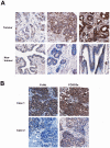
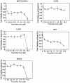
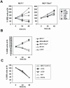
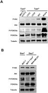
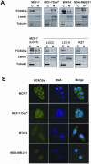
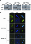
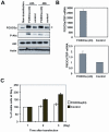
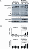
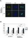
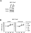
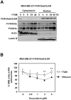
Methodology/principal findings: Examination of FOXO3a and phosphorylated-Akt (P-Akt) expression in breast cancer tissue microarrays showed nuclear FOXO3a was associated with lymph node positivity (p = 0.052), poor prognosis (p = 0.014), and P-Akt expression in invasive ductal carcinoma. Using tamoxifen and doxorubicin-sensitive and -resistant breast cancer cell lines as models, we found that doxorubicin- but not tamoxifen-resistance is associated with nuclear accumulation of FOXO3a, consistent with the finding that sustained nuclear FOXO3a is associated with poor prognosis. We also established that doxorubicin treatment induces proliferation arrest and FOXO3a nuclear relocation in sensitive breast cancer cells. Induction of FOXO3a activity in doxorubicin-sensitive MCF-7 cells was sufficient to promote Akt phosphorylation and arrest cell proliferation. Conversely, knockdown of endogenous FOXO3a expression reduced PI3K/Akt activity. Using MDA-MB-231 cells, in which FOXO3a activity can be induced by 4-hydroxytamoxifen, we showed that FOXO3a induction up-regulates PI3K-Akt activity and enhanced doxorubicin resistance. However FOXO3a induction has little effect on cell proliferation, indicating that FOXO3a or its downstream activity is deregulated in the cytotoxic drug resistant breast cancer cells. Thus, our results suggest that sustained FOXO3a activation can enhance hyperactivation of the PI3K/Akt pathway.
Conclusions/significance: Together these data suggest that lymph node metastasis and poor survival in invasive ductal breast carcinoma are linked to an uncoupling of the Akt-FOXO3a signaling axis. In these breast cancers activated Akt fails to inactivate and re-localize FOXO3a to the cytoplasm, and nuclear-targeted FOXO3a does not induce cell death or cell cycle arrest. As such, sustained nuclear FOXO3a expression in breast cancer may culminate in cancer progression and the development of an aggressive phenotype similar to that observed in cytotoxic chemotherapy resistant breast cancer cell models.
Conflict of interest statement
Figures


Similar articles
-
The forkhead transcription factor FOXO3a increases phosphoinositide-3 kinase/Akt activity in drug-resistant leukemic cells through induction of PIK3CA expression.Mol Cell Biol. 2008 Oct;28(19):5886-98. doi: 10.1128/MCB.01265-07. Epub 2008 Jul 21. Mol Cell Biol. 2008. PMID: 18644865 Free PMC article.
-
The inhibition of activated hepatic stellate cells proliferation by arctigenin through G0/G1 phase cell cycle arrest: persistent p27(Kip1) induction by interfering with PI3K/Akt/FOXO3a signaling pathway.Eur J Pharmacol. 2015 Jan 15;747:71-87. doi: 10.1016/j.ejphar.2014.11.040. Epub 2014 Dec 10. Eur J Pharmacol. 2015. PMID: 25498792
-
Phosphorylation of FOXO3a on Ser-7 by p38 promotes its nuclear localization in response to doxorubicin.J Biol Chem. 2012 Jan 6;287(2):1545-55. doi: 10.1074/jbc.M111.284224. Epub 2011 Nov 29. J Biol Chem. 2012. PMID: 22128155 Free PMC article.
-
Role and regulation of the forkhead transcription factors FOXO3a and FOXM1 in carcinogenesis and drug resistance.Chin J Cancer. 2013 Jul;32(7):365-70. doi: 10.5732/cjc.012.10277. Epub 2013 May 27. Chin J Cancer. 2013. PMID: 23706767 Free PMC article. Review.
-
A new fork for clinical application: targeting forkhead transcription factors in cancer.Clin Cancer Res. 2009 Feb 1;15(3):752-7. doi: 10.1158/1078-0432.CCR-08-0124. Clin Cancer Res. 2009. PMID: 19188143 Free PMC article. Review.
Cited by
-
Increased expression of forkhead box M1 is associated with aggressive phenotype and poor prognosis in estrogen receptor-positive breast cancer.J Korean Med Sci. 2015 Apr;30(4):390-7. doi: 10.3346/jkms.2015.30.4.390. Epub 2015 Mar 19. J Korean Med Sci. 2015. PMID: 25829806 Free PMC article.
-
The prognostic impact of Akt isoforms, PI3K and PTEN related to female steroid hormone receptors in soft tissue sarcomas.J Transl Med. 2011 Nov 22;9:200. doi: 10.1186/1479-5876-9-200. J Transl Med. 2011. PMID: 22107784 Free PMC article.
-
Acidic Fibroblast Growth Factor Promotes Endothelial Progenitor Cells Function via Akt/FOXO3a Pathway.PLoS One. 2015 Jun 10;10(6):e0129665. doi: 10.1371/journal.pone.0129665. eCollection 2015. PLoS One. 2015. PMID: 26061278 Free PMC article.
-
FoxO3a Inhibits Tamoxifen-Resistant Breast Cancer Progression by Inducing Integrin α5 Expression.Cancers (Basel). 2022 Jan 2;14(1):214. doi: 10.3390/cancers14010214. Cancers (Basel). 2022. PMID: 35008379 Free PMC article.
-
Forkhead box K2 modulates epirubicin and paclitaxel sensitivity through FOXO3a in breast cancer.Oncogenesis. 2015 Sep 7;4(9):e167. doi: 10.1038/oncsis.2015.26. Oncogenesis. 2015. PMID: 26344694 Free PMC article.
References
-
- Ali S, Coombes RC. Endocrine-responsive breast cancer and strategies for combating resistance. Nat Rev Cancer. 2002;2:101–112. - PubMed
-
- Elkak AE, Mokbel K. Pure antiestrogens and breast cancer. Curr Med Res Opin. 2001;17:282–289. - PubMed
-
- Yamashita H. Current research topics in endocrine therapy for breast cancer. Int J Clin Oncol. 2008;13:380–383. - PubMed
-
- Goss PE, Muss HB, Ingle JN, Whelan TJ, Wu M. Extended adjuvant endocrine therapy in breast cancer: current status and future directions. Clin Breast Cancer. 2008;8:411–417. - PubMed
-
- Johnston SR. Acquired tamoxifen resistance in human breast cancer—potential mechanisms and clinical implications. Anticancer Drugs. 1997;8:911–930. - PubMed
Publication types
MeSH terms
Substances
Grants and funding
LinkOut - more resources
Full Text Sources
Other Literature Sources
Medical
Research Materials
Miscellaneous

