. 2010 Aug 19;6(8):e1001056.
doi: 10.1371/journal.ppat.1001056.

Enterohemorrhagic E. coli requires N-WASP for efficient type III translocation but not for EspFU-mediated actin pedestal formation

Affiliations

Enterohemorrhagic E. coli requires N-WASP for efficient type III translocation but not for EspFU-mediated actin pedestal formation

Didier Vingadassalom et al. PLoS Pathog. .

Abstract

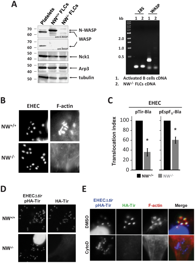
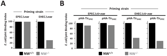
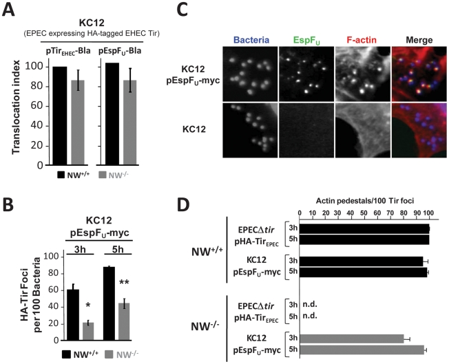
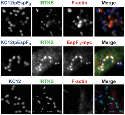
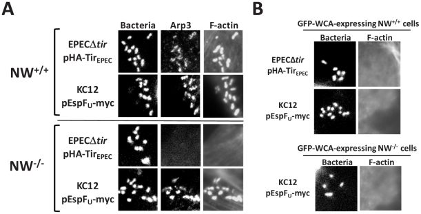
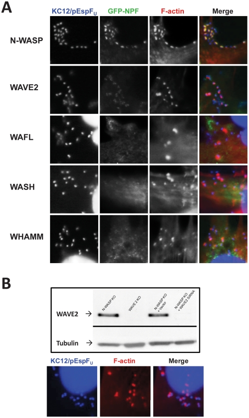
Upon infection of mammalian cells, enterohemorrhagic E. coli (EHEC) O157:H7 utilizes a type III secretion system to translocate the effectors Tir and EspF(U) (aka TccP) that trigger the formation of F-actin-rich 'pedestals' beneath bound bacteria. EspF(U) is localized to the plasma membrane by Tir and binds the nucleation-promoting factor N-WASP, which in turn activates the Arp2/3 actin assembly complex. Although N-WASP has been shown to be required for EHEC pedestal formation, the precise steps in the process that it influences have not been determined. We found that N-WASP and actin assembly promote EHEC-mediated translocation of Tir and EspF(U) into mammalian host cells. When we utilized the related pathogen enteropathogenic E. coli to enhance type III translocation of EHEC Tir and EspF(U), we found surprisingly that actin pedestals were generated on N-WASP-deficient cells. Similar to pedestal formation on wild type cells, Tir and EspF(U) were the only bacterial effectors required for pedestal formation, and the EspF(U) sequences required to interact with N-WASP were found to also be essential to stimulate this alternate actin assembly pathway. In the absence of N-WASP, the Arp2/3 complex was both recruited to sites of bacterial attachment and required for actin assembly. Our results indicate that actin assembly facilitates type III translocation, and reveal that EspF(U), presumably by recruiting an alternate host factor that can signal to the Arp2/3 complex, exhibits remarkable versatility in its strategies for stimulating actin polymerization.


Conflict of interest statement

The authors have declared that no competing interests exist.

Figures

Figure 1
Figure 1. N-WASP-mediated actin assembly facilitates type III translocation of Tir and EspFU from EHEC.
(A) Extracts from platelets, N-WASP positive fibroblast-like cells (“NW+/+ FLCs”) or N-WASP knockout (NW−/−) FLCs were resolved by SDS-PAGE and immunoblotted for N-WASP, WASP, Nck1, Arp3 or tubulin (Left). RT-PCR analysis of WASP mRNA was performed for RNA extracted from NW−/− cells or activated B cells (Right). (B) NW+/+ or NW−/− cells were infected with EHEC and examined after staining with DAPI to localize bacteria and Alexa568-phalloidin to detect F-actin. (C) Translocation of a Tir-Bla or EspFU-Bla fusion by EHEC in NW+/+ and NW−/− cells was measured by detecting cleavage of the β-lactamase FRET reporter CCF2-AM, which results in a change in fluorescent emission of cells from green (absence of detectable Tir-Bla) to blue (presence of Tir-Bla) . Monolayers were infected for 6 hours, incubated with CCF2-AM, and fixed. The percentage of blue cells was scored visually by fluorescent microscopy to determine the translocation index. Shown is the mean ± SD of three experiments; *P<0.05. (D) NW+/+ and NW−/− FLCs were infected with EHECΔtir harboring plasmid pHA-Tir for 5 hours and examined after staining with DAPI to detect bacteria, anti-HA antibody to detect Tir foci and Alexa568-phalloidin for detection of F-actin. (E) DMSO- or cytochalasin D-treated HeLa cells were infected with EHECΔtir harboring plasmid pHA-Tir, fixed, and stained with DAPI to detect bacteria, anti-HA antibody to visualize Tir foci and Alexa568-phalloidin to detect F-actin.
Figure 2
Figure 2. N-WASP is required for efficient intimin-mediated bacterial attachment to the cell surface.
(A) A so-called “prime and challenge” assay was utilized to evaluate Tir/intimin-mediated binding of intimin-expressing E. coli to NW+/+ or NW−/− FLCs. Cells were infected with EPECΔeae or EHECΔeae strains to allow for translocation Tir. After gentamicin treatment to kill EPEC and EHEC, TirEPEC- and TirEHEC-“primed” cells were “challenged” (i.e. infected) for 1 hour, respectively with E. coli K12/pIntEPEC or K12/pIntEHEC that also harbor a plasmid encoding GFP and examined microscopically to detect bound bacteria. The bacterial binding index was defined as the percentage of cells with at least five bound bacteria. (B) NW+/+ or NW−/− cells were infected with EPECΔtir-eae or EHECΔtir-eae harboring either pHA-TirEPEC or pHA-TirEHEC and the bacterial binding index determined as described as above. The experiments described in A and B were performed two times; data from one representative experiment are presented.
Figure 3
Figure 3. TirEHEC and EspFU promote pedestal formation in the absence of N-WASP.
(A) Translocation of the TirEHEC-Bla and EspFU-Bla fusions by KC12 in NW+/+ and NW−/− FLCs was measured 6 hours postinfection in the TEM-1 β-lactamase translocation assay as described in Figure 1. Shown is the mean ± SD of three experiments. (B) NW+/+ and NW−/− cells were infected with KC12/pEspFU-myc, and the percentage of HA-Tir foci associated with bound bacteria was determined at 3 and 5 hours postinfection. Values represent the mean ± SD of three experiments; *P<0.05; **P<0.01. (C) NW+/+ and NW−/− cells were infected with KC12 or KC12/pEspFU-myc and examined after staining with DAPI, anti-myc antibody and Alexa568-phalloidin. (D) NW+/+ or NW−/− cells were infected with EPECΔtir/pHA-TirEPEC or KC12/pEspFU-myc, and then fixed, and stained with DAPI, anti-HA antibody to visualize Tir foci and Alexa568-phalloidin. To quantitate pedestal formation, the percentage of actin pedestals that colocalized with HA-Tir foci-associated bacteria at 3 and 5 hours postinfection was determined visually by fluorescent microscopy (right panel). Data represent the mean ± SD from three experiments. “n.d.”; not detected.
Figure 4
Figure 4. The Tir-EspFU linker protein IRTKS localizes to bacteria even in the absence of N-WASP.
NW−/− FLCs were infected with KC12/pEspFU or KC12, and examined after staining with DAPI to detect bacteria, anti-IRTKS antibody (green) and Alexa568-phalloidin to detect F-actin (red). To detect EspFU-myc, monolayers were treated with rabbit anti-myc antibody and Alexa568-conjugated anti-rabbit antibody (red, middle row).
Figure 5
Figure 5. EspFU is necessary and sufficient to drive N-WASP independent pedestal formation.
(A) NW−/− FLCs ectopically expressing HA-Tir derivatives in which the C-terminal cytoplasmic domain was deleted (TirΔC) or replaced with the C-terminal repeats of EspFU (TirΔC-EspFU-[R1-6]) were challenged with an EPECΔtir strain (which expresses intimin), and stained with DAPI (blue), anti-HA antibody (green) and Alexa568-phalloidin (red). (B) NW−/− FLCs ectopically expressing HA-Tir derivatives in which the C-terminal cytoplasmic domain was replaced with the N-terminal translocation sequence (TirΔC-EspFU-[N]) as a negative control or the C-terminal repeats of EspFU (TirΔC-EspFU-[R1-6]) were challenged with E. coli/pInt to cluster the fusion protein, and treated as in A.
Figure 6
Figure 6. The Arp2/3 complex is critical for actin pedestal formation on N-WASP-deficient cells.
(A) NW+/+ or NW−/− FLCs were infected with KC12/pEspFU or EPECΔtir/pHA-TirEPEC, fixed, and stained with DAPI to detect bacteria, anti-Arp3 antibody to visualize the Arp2/3 complex and Alexa568-phalloidin to detect F-actin. (B) Transfected NW+/+ or NW−/− cells expressing GFP fused to the WCA domain of N-WASP (GFP-WCA) were infected with KC12/pEspFU or EPECΔtir/pHA-TirEPEC. Monolayers were stained with DAPI and Alexa568-phalloidin, and transfected cells were identified by GFP fluorescence. In two independent experiments, expression of the GFP-WCA fusion protein strongly inhibited pedestal formation.
Figure 7
Figure 7. WASP/WAVE family members that are not involved in pedestal formation.
(A) NW−/− fibroblast-like cells ectopically expressing GFP fusions to WASP/WAVE family members N-WASP, WAVE2, WASH, or WHAMM, or to WAFL, were infected with EPEC KC12/pEspFU, and examined after staining with DAPI to detect bacteria (blue) and Alexa568-phalloidin to visualize F-actin (red). (B) Genetic depletion of WAVE2 in N-WASP knockout cells does not affect pedestal formation. Extracts from NW−/− cells, WAVE2−/− cells, or NW−/− cells harboring control vector or one expressing a WAVE2 siRNA, were resolved by SDS-PAGE and immunoblotted for WAVE2 or tubulin, as a loading control (Top panel). NW−/−/WAVE2 knockdown cells were infected with KC12/pEspFU, and examined after staining with DAPI to detect bacteria and Alexa568-phalloidin to visualize F-actin (Bottom panel).
Figure 8
Figure 8. Pedestal formation on N-WASP-deficient cells requires multiple EspFU repeats.
(A) NW−/− cells (shaded bars) ectopically expressing Tir-EspFU fusion derivatives were treated with anti-Tir antibodies and S. aureus particles to promote membrane clustering. Monolayers were stained with anti-HA antibody to identify both transfected cells and S. aureus (which binds the fluorescent antibodies) and with Alexa568-phalloidin to detect F-actin. The pedestal formation indices were determined by calculating the percentage of transfected cells harboring five or more S. aureus particles associated with actin pedestals. These experiments were performed in parallel with NW+/+ cells (solid bars), the results of which were previously published and are shown here for comparison. Data represent the mean ± SD from three experiments. (B) NW−/− (shaded bars) ectopically expressing GFP-EspFU fusion derivatives were infected with EPEC KC12 and monolayers stained with DAPI to identify attached bacteria, anti-myc antibody to enhance detection of GFP-EspFU fusion and Alexa568-phalloidin to detect F-actin. The pedestal formation indices were determined by calculating the percentage of transfected cells harboring five or more actin pedestals. These experiments were performed in parallel with NW+/+ (solid bars) cells, the results of which were previously published and are shown here for comparison. Data represent the mean ± SD from three experiments. “n.d.”; not detected.
Figure 9
Figure 9. The WASP/N-WASP-binding region of EspFU is required for N-WASP-independent actin pedestal formation.
(A) Sequence alignment of an EspFU repeat and the corresponding VLL/AAA mutant. The WASP/N-WASP-binding α-helix is colored in blue. (B) NW−/− FLCs ectopically expressing HA-Tir-EspFU-2RWT or HA-Tir-EspFU-2RVLL/AAA fusions were challenged with an EPECΔtir strain (which expresses intimin), and stained with DAPI (blue), anti-HA antibody (green) and Alexa568-phalloidin (red).

Similar articles

Cited by

References

    1. Kaper JB, Nataro JP, Mobley HL. Pathogenic Escherichia coli. Nat Rev Microbiol. 2004;2:123–140. - PubMed
    1. Deng W, Vallance BA, Li Y, Puente JL, Finlay BB. Citrobacter rodentium translocated intimin receptor (Tir) is an essential virulence factor needed for actin condensation, intestinal colonization and colonic hyperplasia in mice. Mol Microbiol. 2003;48:95–115. - PubMed
    1. Marches O, Nougayrede JP, Boullier S, Mainil J, Charlier G, et al. Role of tir and intimin in the virulence of rabbit enteropathogenic Escherichia coli serotype O103:H2. Infect Immun. 2000;68:2171–2182. - PMC - PubMed
    1. Ritchie JM, Thorpe CM, Rogers AB, Waldor MK. Critical roles for stx2, eae, and tir in enterohemorrhagic Escherichia coli-induced diarrhea and intestinal inflammation in infant rabbits. Infect Immun. 2003;71:7129–7139. - PMC - PubMed
    1. Tacket CO, Sztein MB, Losonsky G, Abe A, Finlay BB, et al. Role of EspB in experimental human enteropathogenic Escherichia coli infection. Infect Immun. 2000;68:3689–3695. - PMC - PubMed

Publication types

MeSH terms