. 2010 Aug 19;5(8):e12270.
doi: 10.1371/journal.pone.0012270.

Single-molecule three-color FRET with both negligible spectral overlap and long observation time

Affiliations

Single-molecule three-color FRET with both negligible spectral overlap and long observation time

Sanghwa Lee et al. PLoS One. .

Abstract

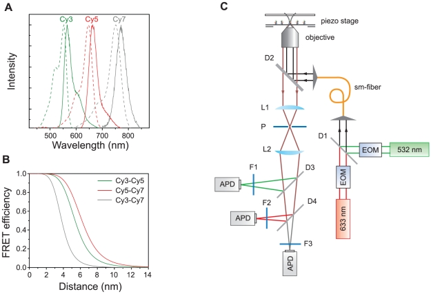
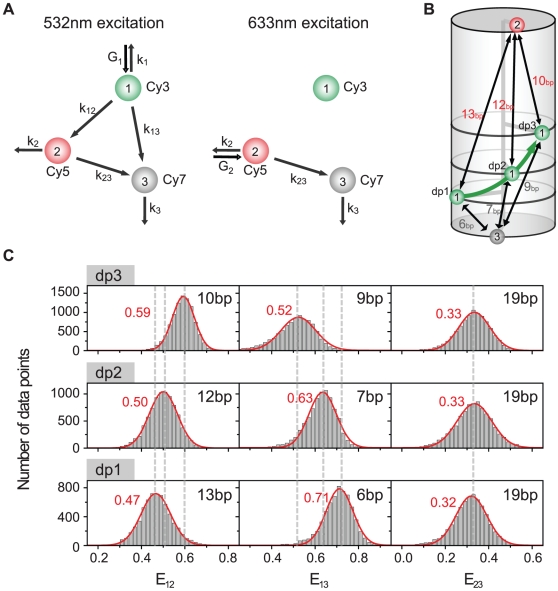
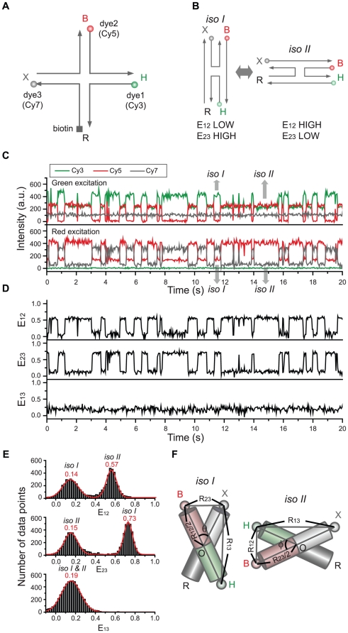
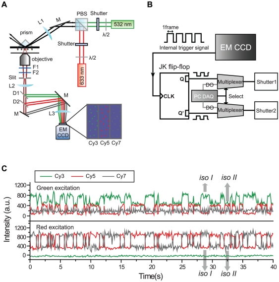
Full understanding of complex biological interactions frequently requires multi-color detection capability in doing single-molecule fluorescence resonance energy transfer (FRET) experiments. Existing single-molecule three-color FRET techniques, however, suffer from severe photobleaching of Alexa 488, or its alternative dyes, and have been limitedly used for kinetics studies. In this work, we developed a single-molecule three-color FRET technique based on the Cy3-Cy5-Cy7 dye trio, thus providing enhanced observation time and improved data quality. Because the absorption spectra of three fluorophores are well separated, real-time monitoring of three FRET efficiencies was possible by incorporating the alternating laser excitation (ALEX) technique both in confocal microscopy and in total-internal-reflection fluorescence (TIRF) microscopy.


Conflict of interest statement

Competing Interests: The authors have declared that no competing interests exist.

Figures

Figure 1
Figure 1. Dye selection and a confocal setup.
(a) Normalized emission (solid lines) and absorption (dash lines) spectra of Cy3 (green), Cy5 (red) and Cy7 (gray). (b) FRET efficiencies of the three FRET pairs as a function of inter-dye distances. R0 values of Cy3-Cy5 (green), Cy5-Cy7 (red) and Cy3-Cy7 (gray) pairs were calculated as 5.4-nm, 6.2-nm, and 3.8-nm, respectively. (c) A schematic diagram of ALEX three-color confocal setup. The setup was built based on an inverted microscope (TE2000-U, Nikon, Tokyo, Japan) equipped with a three dimensional piezo-stage (LP-100, MadCityLabs, Madison, WI). Two excitation lasers, a diode-pump solid state laser (532-nm, Excelsior-CDRH, Spectra-Physics, Santa Clara, CA) and HeNe laser (633-nm, HRP050, Thorlabs, Newton, NJ), were alternatively switched on and off by using electro-optic modulators (EOM, 350-50, Conoptics, Danbury, CT). To make sure that the two excitation lasers excite the same molecule, they were coupled into a single-mode fiber (460HP, Thorlabs). An oil-immersion objective (UPLSAPO 100×, Olympus, Tokyo, Japan) was used for both the excitation of molecules and the collection of fluorescence signals. The fluorescence signals are measured by using avalanche photo diodes (APD, SPCM-AQRH-14, Perkin Elmer, Wellesley, MA). The identities of other optics are: D1, a dichroic mirror (z532bcm, Chroma, Rockingham, VT); D2, dichroic mirror (z532/633rpc, Chroma); P, pinhole (P75S, Thorlabs); L1 and L2, lens (LAO-90.0-25.0/078, CVI, Irvine, CA); D3, dichroic mirror (640dcxr, Chroma); D4, dichroic mirror (740dcxr, Chroma); F1, bandpass filter (HQ580/60m-2p, Chroma); F2, bandpass filter (HQ680/60m-2p, Chroma); F3, bandpass filter (HQ790/80m, Chroma).
Figure 2
Figure 2. Determination of three FRET efficiencies.
(a) An interaction diagram of three dyes upon 532-nm and 633-nm excitations. The identities of symbols used here are explained in the text. (b) Triply-labeled DNA duplexes. The positions of Cy5 and Cy7 are the same, but the labeling position of Cy3 is different in dp1, dp2, dp3. (c) FRET efficiency histograms of the three FRET pairs for each DNA duplex.
Figure 3
Figure 3. Conformational dynamics of the triply-labeled Holliday junction observed in the confocal setup.
(a) Dye labeling scheme of the Holliday junction. (b) Dynamics of the Holliday junction between two conformers, isoI and isoII. (c) Typical fluorescence intensity time traces of Cy3 (green lines), Cy5 (red lines) and Cy7 (gray lines) of the Holliday junction upon 532-nm excitation (upper graphs) or 633-nm excitation (lower graphs). Two excitation lasers were alternatively switched on and off every 20 millisecond synchronously with data binning. The experiments were performed at room temperature in 10 mM Tris-HCl (pH 8.0) with 50 mM Mg2+. (d) FRET efficiency time traces calculated from the data in (c). (e) Three inter-dye FRET histograms of the Holliday junction. The histograms were made from more than 20 molecules. (f) Diagram for the determination of inter-duplex angles of isoI and isoII.
Figure 4
Figure 4. Conformational dynamics of the triply-labeled Holliday junction observed in the TIRF setup.
(a) A schematic diagram of ALEX three-color FRET setup in TIRF microscopy. Then setup was built based on an inverted microscope (IX71, Olympus). For switching purposes, mechanical shutters (LS-3, Uniblitz, Rochester, NY) were used. The fluorescence signals of the three dyes were collected by a water-immersion objective (UPlanSApo 60×, Olympus) and focused on different areas of an electron-multiflier charge coupled device (EM-CCD, iXon+ DU897BV, Andor Technology, Belfast, UK). The identities of other optics are: F1, notch filter (NF03-633E-25, Semrock, Rochester, NY); F2, long-pass filter (LP03-532RU-25, Semrock); D1, dichroic mirror (635dcxr, Chroma); D2, dichroic mirror (740dcxr, Chroma); L1, lens (LA1986-A, Thorlabs); L2, lens (LAO-120.0-40.0/066, CVI); L3, lens (LAO-260.1-50.0/066, CVI); M, mirror (BB01-E02, Thorlabs). (b) A circuit diagram for synchronization of laser switching and image acquisition. The internal trigger line of the EM-CCD is connected to the clock line of a JK flip-flop (74LS112, Motorola, Schaumburg, IL), and the JK flip-flop generates two complementary pulse trains, Q and Q′, which are used to alternately switch two excitation lasers synchronously. A multiplexer is used to select either a data acquisition board (DAQ PCI-6503, NI) or EM-CCD as a source of shutter control signal. (c) Fluorescence intensity time traces of the Holliday junction observed in TIRF microscope with 50-ms bin time. Experiments were performed at room temperature in 10 mM Tris-HCl (pH 8.0) with 200 mM Mg2+.

References

    1. Ha T, Enderle T, Ogletree DF, Chemla DS, Selvin PR, et al. Probing the interaction between two single molecules: fluorescence resonance energy transfer between a single donor and a single acceptor. Proc Natl Acad Sci U S A. 1996;93:6264–6268. - PMC - PubMed
    1. Ha T. Single-molecule fluorescence resonance energy transfer. Methods. 2001;25:78–86. - PubMed
    1. Hohng S, Zhou R, Nahas MK, Yu J, Schulten K, et al. Fluorescence-force spectroscopy maps two-dimensional reaction landscape of the Holliday junction. Science. 2007;318:279–283. - PMC - PubMed
    1. Tarsa PB, Brau RR, Barch M, Ferrer JM, Freyzon Y, et al. Detecting force-induced molecular transitions with fluorescence resonant energy transfer. Angew Chem Int Ed. 2007;46:1999–2001. - PMC - PubMed
    1. Shroff H, Reinhard BM, Siu M, Agarwal H, Spakowitz A, et al. Biocompatible force sensor with optical readout and dimensions of 6nm3. Nano Lett. 2005;5:1509–1514. - PubMed

Publication types