. 2010 Aug 19;6(8):e1001050.
doi: 10.1371/journal.ppat.1001050.

Functional characterisation and drug target validation of a mitotic kinesin-13 in Trypanosoma brucei

Affiliations

Functional characterisation and drug target validation of a mitotic kinesin-13 in Trypanosoma brucei

Kuan Yoow Chan et al. PLoS Pathog. .

Abstract

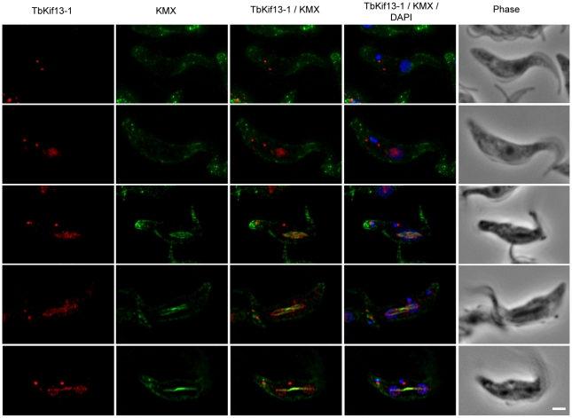
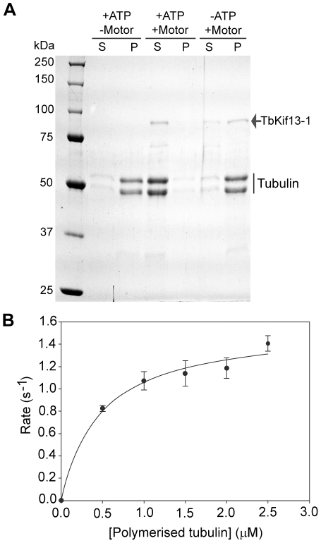
Mitotic kinesins are essential for faithful chromosome segregation and cell proliferation. Therefore, in humans, kinesin motor proteins have been identified as anti-cancer drug targets and small molecule inhibitors are now tested in clinical studies. Phylogenetic analyses have assigned five of the approximately fifty kinesin motor proteins coded by Trypanosoma brucei genome to the Kinesin-13 family. Kinesins of this family have unusual biochemical properties because they do not transport cargo along microtubules but are able to depolymerise microtubules at their ends, therefore contributing to the regulation of microtubule length. In other eukaryotic genomes sequenced to date, only between one and three Kinesin-13s are present. We have used immunolocalisation, RNAi-mediated protein depletion, biochemical in vitro assays and a mouse model of infection to study the single mitotic Kinesin-13 in T. brucei. Subcellular localisation of all five T. brucei Kinesin-13s revealed distinct distributions, indicating that the expansion of this kinesin family in kinetoplastids is accompanied by functional diversification. Only a single kinesin (TbKif13-1) has a nuclear localisation. Using active, recombinant TbKif13-1 in in vitro assays we experimentally confirm the depolymerising properties of this kinesin. We analyse the biological function of TbKif13-1 by RNAi-mediated protein depletion and show its central role in regulating spindle assembly during mitosis. Absence of the protein leads to abnormally long and bent mitotic spindles, causing chromosome mis-segregation and cell death. RNAi-depletion in a mouse model of infection completely prevents infection with the parasite. Given its essential role in mitosis, proliferation and survival of the parasite and the availability of a simple in vitro activity assay, TbKif13-1 has been identified as an excellent potential drug target.


Conflict of interest statement

The authors have declared that no competing interests exist.

Figures

Figure 1
Figure 1. Immunolocalisation of trypanosome Kinesin-13 members.
Fluorescence images of procyclic cells stained with anti-Kinesin13 antibodies (red). Nuclear and kinetoplast DNA has been counterstained with DAPI (blue). The corresponding phase contrast images are shown in the right panel. Immunolocalisation of endogenous TbKif13-1, TbKif13-3 and TbKif13-4 was achieved using polyclonal antibodies raised against protein fragments specific to each of the kinesins. The immunolocalisation of TbKif13-2 and TbKif13-5 was done by overexpressing a C-terminal cmyc-tagged recombinant version of the corresponding kinesin employing a tet-inducible expression system. An anti-myc monoclonal antibody was used for the detection of the epitope-tagged proteins. Bar, 2 µm.
Figure 2
Figure 2. Cell cycle dependent localisation of TbKif13-1.
Cellular localisation of TbKif13-1 (red) during the cell cycle. The position of a cell in the cell cycle was assessed by its number of kinetoplasts and nuclei. 1K1N cells are in interphase, 2K1N are either in late G2 or early mitosis and 2K2N cells are in mitosis. The mitotic spindle was stained with the anti-tubulin antibody KMX (green). Nuclear and kinetoplast DNA is stained with DAPI (blue). Bar, 2µm.
Figure 3
Figure 3. TbKif13-1 is an essential protein in trypanosomes.
(A) Western blot on whole cell lysates of bloodstream (BSF) and procyclic (PCF) TbKif13-1 RNAi-depleted cells, either non-induced (−) or induced (+) with doxycycline for 2 days. The Coomassie staining serves as a loading control. (B) A cumulative logarithmic growth curve comparing the growth rates of induced and non-induced (control) procyclic and bloodstream RNAi cells. (C) Fluorescence images of procyclic non-induced (−Dox) or induced (+Dox) for two days. RNAi-mediated depletion results in the loss of nuclear staining, but leaves the cytoplasmic dot-like structures unaffected. The position of the kinetoplasts is indicated by arrowheads. Bar, 2 µm.
Figure 4
Figure 4. Depletion of TbKif13-1 results in abnormal nucleus and kinetoplast numbers.
(A) Analysis of the numbers of kinetoplasts (K) and nuclei (N) in bloodstream (BSF) and procyclic (PCF) cells. (K*N*) denotes cells with morphologically abnormal nuclei and/or >2 kinetoplast. At least 100 cells were counted for each time point. (B) Phase contrast and the corresponding DAPI stained fluorescence images of non-induced (−Dox) and induced (+Dox) cells. Bar, 2 µm. (C) Flow cytometry analysis of non-induced (control) and induced cells treated with doxycycline for one or two days.
Figure 5
Figure 5. TbKif13-1 depletion results in chromosome segregation defects.
Combined immunofluorescence and fluorescent in situ hybridisation (FISH) of procyclic cells using antibody KMX (green) to visualise the mitotic spindle and the 177bp-repeat probe to visualise minichromosomes (red). Shown are representative examples of non-induced and induced TbKif13-1 RNAi cells. Total DNA was stained with DAPI (blue). Phase contrast images of cells are shown in the right panel. Bar, 2 µm.
Figure 6
Figure 6. TbKif13-1 depletion causes an extended spindle phenotype.
Bloodstream (BSF) and procyclic (PCF) T. brucei cells depleted of TbKif13-1 using RNAi were stained with KMX (green) to visualise the mitotic spindle and DAPI to stain nuclear and kinetoplast DNA (blue). Non-induced cells are shown for both life cycle stages to illustrate normal spindle appearance during early (BSF, upper panel, rhomboid spindle) and late mitosis (PCF, upper panel, bifurcated spindle). Note the appearance of bent spindles in late mitosis in BSF and PCF cells (traced with white dots). Bar, 2 µm.
Figure 7
Figure 7. TbKif13-1 depletion results in protrusions of the nuclear envelope.
(A) Procyclic TbKif13-1 depleted cells were stained with NUP, an antibody specific to the nuclear envelope. Bar, 2 µm. (B) An EM image of a nuclear cross section of a TbKif13-1 depleted cell. The cell is in mitosis as indicated by the presence of two electron dense regions within the nucleus representing the dividing nucleolus, thus defining the geometry of a dividing nucleus. A protrusion of the nuclear envelope is marked by a white arrow. Several microtubules extend into these protrusions (white arrowheads). Bar 0.5 µm.
Figure 8
Figure 8. In vitro biochemical analysis of recombinant TbKif13-1.
(A) Microtubule depolymerisation assay of TbKif13-1 in the presence (+) or absence (−) of ATP/kinesin. S, supernatant fraction; P, pellet fraction. (B) ATPase activity of TbKif13-1 is stimulated by microtubules. Each data point represents three independent measurements. A curve of best fit was drawn using the Michaelis-Menten equation in SigmaPlot V11.0.

Similar articles

Cited by

References

    1. Dacks JB, Walker G, Field MC. Implications of the new eukaryotic systematics for parasitologists. Parasitol Int. 2008;57:97–104. - PubMed
    1. Gull K. The cytoskeleton of trypanosomatid parasites. Annu Rev Microbiol. 1999;53:629–655. - PubMed
    1. McKean PG, Vaughan S, Gull K. The extended tubulin superfamily. J Cell Sci. 2001;114:2723–2733. - PubMed
    1. Dawe HR, Farr H, Portman N, Shaw MK, Gull K. The Parkin co-regulated gene product, PACRG, is an evolutionarily conserved axonemal protein that functions in outer-doublet microtubule morphogenesis. J Cell Sci 2005 - PubMed
    1. Dawe HR, Shaw MK, Farr H, Gull K. The hydrocephalus inducing gene product, Hydin, positions axonemal central pair microtubules. BMC Biol. 2007;5:33. - PMC - PubMed

Publication types

MeSH terms

LinkOut - more resources