. 2010 Aug 19;6(8):e1001052.
doi: 10.1371/journal.ppat.1001052.

Damaged intestinal epithelial integrity linked to microbial translocation in pathogenic simian immunodeficiency virus infections

Affiliations

Damaged intestinal epithelial integrity linked to microbial translocation in pathogenic simian immunodeficiency virus infections

Jacob D Estes et al. PLoS Pathog. .

Abstract

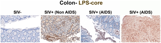
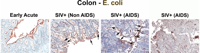
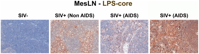
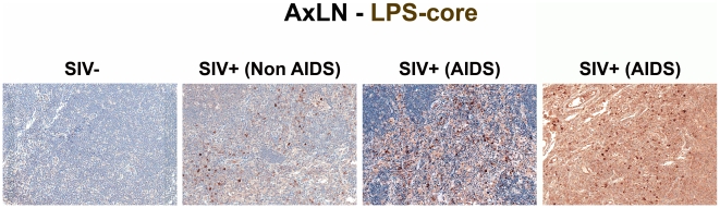
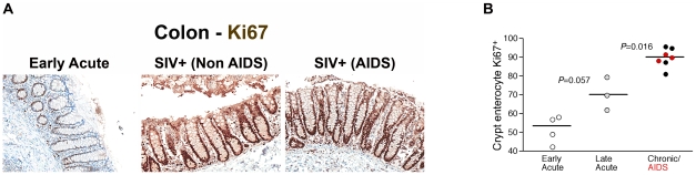
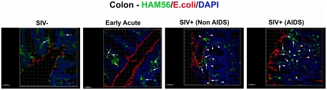
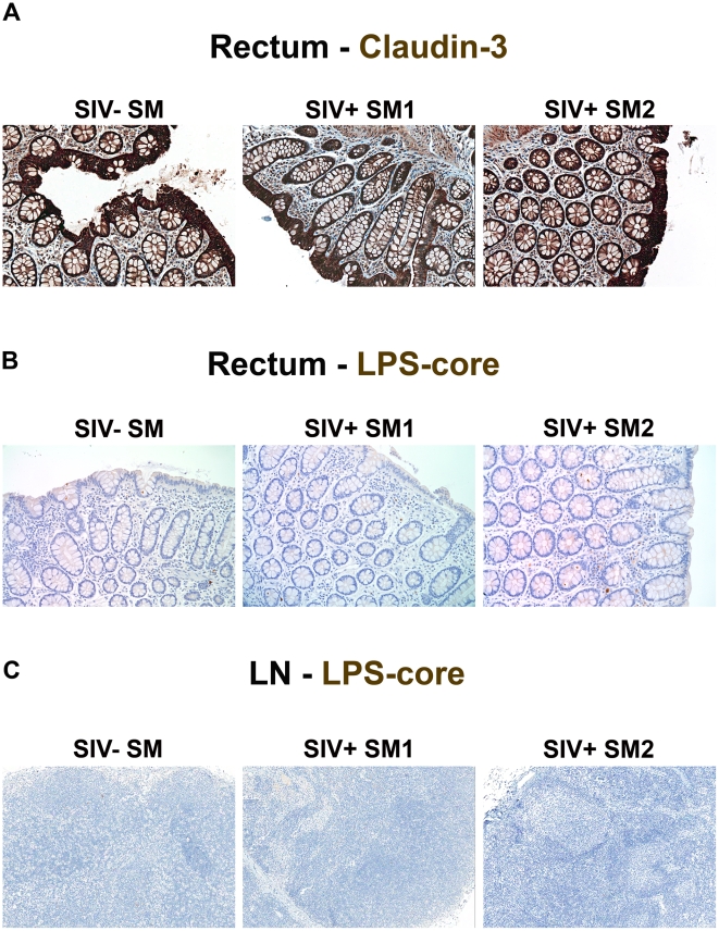
The chronic phase of HIV infection is marked by pathological activation of the immune system, the extent of which better predicts disease progression than either plasma viral load or CD4(+) T cell count. Recently, translocation of microbial products from the gastrointestinal tract has been proposed as an underlying cause of this immune activation, based on indirect evidence including the detection of microbial products and specific immune responses in the plasma of chronically HIV-infected humans or SIV-infected Asian macaques. We analyzed tissues from SIV-infected rhesus macaques (RMs) to provide direct in situ evidence for translocation of microbial constituents from the lumen of the intestine into the lamina propria and to draining and peripheral lymph nodes and liver, accompanied by local immune responses in affected tissues. In chronically SIV-infected RMs this translocation is associated with breakdown of the integrity of the epithelial barrier of the gastrointestinal (GI) tract and apparent inability of lamina propria macrophages to effectively phagocytose translocated microbial constituents. By contrast, in the chronic phase of SIV infection in sooty mangabeys, we found no evidence of epithelial barrier breakdown, no increased microbial translocation and no pathological immune activation. Because immune activation is characteristic of the chronic phase of progressive HIV/SIV infections, these findings suggest that increased microbial translocation from the GI tract, in excess of capacity to clear the translocated microbial constituents, helps drive pathological immune activation. Novel therapeutic approaches to inhibit microbial translocation and/or attenuate chronic immune activation in HIV-infected individuals may complement treatments aimed at direct suppression of viral replication.


Conflict of interest statement

The authors have declared that no competing interests exist.

Figures

Figure 1
Figure 1. Identification of microbial translocation in large bowel of chronically SIV+ RMs.
Representative images (200×) of unselected colon sections from SIV-uninfected and chronically SIV-infected RMs (Non-AIDS and AIDS) stained for LPS-core antigen (brown). The images show the array of microbial translocation and damage seen in our cohort that ranged from negligible (SIV- RMs) to regions of epithelial loss in our SIV+ Non-AIDS RMs resulting in luminal content in direct contact with the LP and increased frequency of LPS+ cells within the LP to more extensive epithelial damage in our SIV+ AIDS RMs that extended to overt ulcerations that showed significant microbial product infiltration throughout these regions; features which were absent from our SIV-uninfected RMs. Arrows point to regions of epithelial discontinuities and microbial translocation into the LP.
Figure 2
Figure 2. Identification of microbial translocation in large bowel of chronically SIV+ RMs.
Representative images (200×) of colon stained with a polyclonal antibody against E. coli (brown) in SIV early acute infected (4 dpi) and chronically SIV-infected RMs (Non-AIDS and AIDS). Note the small regions of epithelial discontinuities in the SIV+ Non-AIDS RMs resulting in luminal E. coli + content in direct contact with the LP and increased frequency of E. coli + cells within the LP. Sections from two SIV+ AIDS RMs show the range of epithelial damage with associated microbial product infiltration, findings frequently seen in RMs with AIDS. Arrows point to regions of epithelial discontinuities and microbial translocation into the LP.
Figure 3
Figure 3. Identification of microbial translocation in gut draining MesLN of chronically SIV+ RMs.
Representative images (200×) of the paracortical T-cell zone of MesLN stained for LPS-core antigen (brown). Note the more extensive accumulation of microbial products within the T cell zone, the T-cell inductive parenchyma of the lymphatic tissue with progressively more severe SIV infection.
Figure 4
Figure 4. Identification of systemic microbial translocation in distant AxLN of chronically SIV+ RMs.
Representative images (200×) of the paracortex of AxLN stained for LPS-core antigen (brown). Note the increased accumulation of microbial products within the paracortex, germinal centers, and medullary chords of the lymphatic tissue, with progressive SIV infection in chronically SIV+ RMs, but the paucity of LPS staining seen in SIV-uninfected RMs.
Figure 5
Figure 5. Identification of microbial translocation in the liver of chronically SIV+ RMs.
Representative images (200×) of liver stained forE. coli constituents (brown). Note the increasing accumulation of microbial products near and surrounding the portal tract in chronically SIV+ RMs with more severe disease but lack of microbial constituent staining seen in SIV-uninfected RMs.
Figure 6
Figure 6. Quantitative image analysis of microbial translocation.
(A) Levels of LPS within the LP of the colon of SIV-uninfected (white squares), early acute (white circles), late acute (gray circles), chronic Non-AIDS (black circles) and chronic AIDS (red circles) RMs. Mann-Whitney U test performed between the chronically infected (both Non-AIDS and AIDS) and early acute RMs and between late acute and early acute RMs. (B) Correlation between LPS levels within the LP of the colon and the mesenteric LN for animals in which we had obtained both colon and MesLN samples. Significance determined using Spearman's rank correlation coefficient and associated p-value shown. (C) Correlation between LPS levels within MesLN and AxLN for animals in which we had obtained both MesLN and AxLN samples. Significance determined using Spearman's rank correlation coefficient and associated P-value shown.
Figure 7
Figure 7. Damage to the integrity of the epithelial barrier is associated with infiltration of microbial products into the lamina propria.
(A) Representative images (200×) of colon stained for the tight junction protein claudin-3 (brown) as a marker for intact epithelial barrier. Note the multifocal regions of the epithelial barrier that are missing in SIV+ RMs, allowing direct contact of luminal contents with the LP. (B) Confocal immunofluorescent 3-D images of colon (400×) stained for cytokeratin (epithelial cells; green), E. coli (red) and DAPI (nuclei; blue). Discontinuities in the epithelial barrier allowing translocation of microbial products into the LP are present in late acute SIV+ RMs (14 dpi) and progressively more severe in chronic Non-AIDS and chronic AIDS RMs (white arrows point to epithelial discontinuities and translocated microbial products). Scale bars  = 40 µm.
Figure 8
Figure 8. Damage to the integrity of the epithelial barrier correlates with microbial translocation.
(A) Quantitative image analysis showing the proportion of the epithelial lining in the large bowel that is damaged (discontinuous) in SIV-uninfected (white squares), early acute (white circles), late acute (gray circles), chronic Non-AIDS (black circles) and chronic AIDS (red circles) RMs. Mann-Whitney U test performed between the chronically infected (both Non-AIDS and AIDS) and early acute RMs and between chronically infected (both Non-AIDS and AIDS) and SIV- RMs. (B) Statistically significant positive correlation between the proportion of epithelial damage and the level of LPS within the LP of the colon. Significance determined using Spearman's rank correlation coefficient and associated P-value shown.
Figure 9
Figure 9. Damage to the integrity of the epithelial barrier is associated with increased enterocyte proliferation.
(A) Representative images (200×) of colon stained for the proliferative nuclear protein Ki67 (brown). Note progressive change in frequency and location of Ki67+ enterocytes, from few cells at the base of crypts during early acute infection (4 dpi), with full involvement of crypts with proliferating cells in chronically infected Non-AIDS and AIDS animals. (B) Quantitative image analysis of Ki67 expression by colonic enterocytes in early acute (white circles), late acute (gray circles), chronic Non-AIDS (black circles) and chronic AIDS (red circles) RMs demonstrates the statistically significant increase in enterocyte proliferation in chronic infection. Mann-Whitney U test performed between the chronically infected (both Non-AIDS and AIDS) and late acute RMs and between early acute and late acute RMs.
Figure 10
Figure 10. Microbial products are differentially phagocytosed in acutely and chronically SIV-infected RMs.
Confocal immunofluorescent 3-D images of colon (400×) stained for HAM56 (macrophages; green), E. coli (red) and DAPI (nuclei; blue) showing the progressive decrease in the fraction of microbial products (E. coli, red) internalized by macrophages (green, highlighted by white arrows), and an increase in the proportion that were not cell associated with progressively more severe SIV infection. For specimens obtained from animals with later stages of infection, the presence of abundant HAM56+ macrophages (green) without internalized E. coli constituents (red) (highlighted by white arrow heads), despite extensive E. coli constituents present in the LP near macrophages, suggests that macrophages in chronic SIV+ RMs become less able to efficiently clear microbial products translocated into the LP. Rare microbial constituents are present in SIV-uninfected RMs but are virtually always found within macrophages. Scale bars  = 40 µm.
Figure 11
Figure 11. Absence of structural epithelial damage and microbial translocation in non-pathogenic infection of SMs.
(A) Representative images (200×) of rectum stained for the tight junction protein claudin-3 (brown) show the complete maintenance of the epithelial barrier in SIVsmm-uninfected and SIVsmm-infected SMs. Representative images (200×) of (B) rectum and (C) peripheral lymph nodes (100×) stained for LPS-core antigen (brown) shows the absence of microbial translocation in SIVsmm-uninfected and SIVsmm-infected SMs.

References

    1. Lane HC. Polyclonal activation of B cells in homosexual men. New Engl J Med. 1984;31:536–537. - PubMed
    1. Hellerstein M, Hanley MB, Cesar D, Siler S, Papageorgopoulos C, et al. Directly measured kinetics of circulating T lymphocytes in normal and HIV-1-infected humans. Nat Med. 1999;5:83–89. - PubMed
    1. Hazenberg MD, Otto SA, van Benthem BH, Roos MT, Coutinho RA, et al. Persistent immune activation in HIV-1 infection is associated with progression to AIDS. Aids. 2003;17:1881–1888. - PubMed
    1. Valdez H, Lederman MM. Cytokines and cytokine therapies in HIV infection. AIDS Clin Rev. 1997. pp. 187–228. - PubMed
    1. Giorgi JV, Hultin LE, McKeating JA, Johnson TD, Owens B, et al. Shorter survival in advanced human immunodeficiency virus type 1 infection is more closely associated with T lymphocyte activation than with plasma virus burden or virus chemokine coreceptor usage. J Infect Dis. 1999;179:859–870. - PubMed

MeSH terms